Acquiring comprehensive 3D images of specimens is essential for performing extensive studies in several applications. Doing so using non-destructive procedures such as X-ray computed tomography (X-ray CT) limits resolution. Destructive approaches, which function based on sequential delayering and scanning of the sample, confront a compromise between throughput and resolution.
Despite its high precision, USING a focused ion beam (FIB) for delayering has a low throughput. Traditional FIBs have alternatives, such as gas-assisted, high-current, and ionized source FIBs. However, these techniques sacrifice precision for speed and need days to remove one cubic mm of material.
Other problems of FIB include high cost and vacuuming, curtaining effects, and charging aberrations for nonconductive substances. However, different known delayering approaches with the potential for larger throughputs have accuracy and controllability difficulties.
On the other hand, mechanical technologies that may enable quick delayering have poor accuracy and risk sample integrity. Another shortcoming of mechanical procedures is the thermal and dynamic stress imposed on the sample, which is detrimental to the sample’s integrity in certain situations.
Potential of Femtosecond Laser Ablation in 3D imaging
As a substitute to the usual mechanical and FIB delayering techniques, femtosecond laser ablation, which produces low to negligible heat-affected zones (HAZs), can be considered.
Rapid material removal efficiencies distinguish laser ablation from FIB in terms of throughput performance. Unlike mechanical techniques, laser ablation does not need rigorous sample preparation processes, does not compromise the sample's integrity, and is material-independent.
Challenges Associated with Femtosecond Laser Ablation
In standard 3D high-resolution volumetric imaging, the revealed layer of the sample at each phase has a flat topography; this assumption is employed when constructing the 3D image by layering the 2D layers.
Owing to variation in the laser ablation rate of various materials, obtaining flat cuts, which are the foundation for acquiring tomographic images using the standard method, causes obstacles.
Variations in the engagement of the laser beam with diverse chemical contents in a multi-material specimen can result in a depth of cut that varies significantly throughout the sample. This effect may significantly affect the final 3D image.
The re-deposition of laser ablation material onto the workpiece is an additional critical challenge posed by lasers, which might substantially affect the final 3D image quality.
Strategy to Enhance Femtosecond Laser Ablation for Rapid 3D imaging
In this study, the researchers suggested using confocal microscopy to overcome the difficulties of generating flat layers.
Instead of focusing on images of 2D layers which may not be flat, this technique incorporates confocal microscopy to obtain a depth map of the subjected layer of the specimen at each stage of the procedure. The team used this information in the 3D image reconstruction. The depth map was also utilized for further lasering design.
Sections cut deeper than a predetermined threshold were omitted from the subsequent lasering process in an attempt, known as masking, to keep the height variation throughout the region of interest within a predefined limit. As a result, the confocal images of the revealed layers will be able to capture the full area of interest.
Key Features of Employing Femtosecond Laser Ablation with Confocal Microscopy
Automatic material characterization and 3D image recognition are significant by-products of Femtosecond Laser Ablation with Confocal Microscopy. The suggested approach takes advantage of the disparity in the ablation rates of various materials when identical lasering parameters are used. Finally, incorporated computer vision algorithms guarantee that the procedure is automated.
Research Findings
The researchers employed femtosecond laser ablation with confocal microscopy on a printed circuit board (PCB). This study presents the outcomes of applying the suggested approach on a PCB to acquire an image with a total height range of approximately 700 µm. It took around 20–30 hours to complete the entire process, which was faster than FIB and other mechanical procedures.
The findings presented by the researchers indicated that the 3D image produced by the suggested technology had a higher information density. First, the optical/confocal 3D image holds all the details that can be extracted from the X-ray CT image.
The resultant optical/confocal 3D picture contains color features, which facilitate the differentiation of various material compositions.
Lastly, the X-ray CT images lack the high-resolution optical evidence from the glass fiber material provided by the suggested approach. Figure 1 illustrates a few XY-plane segments of the 3D PCB image, alongside matching X-ray CT images.
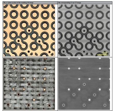
Figure 1. Illustration of several XY-plane segments of the 3D PCB image, alongside matching X-ray CT images. Image Credit: Phoulady, A., et al.
Using Femtosecond Laser Ablation for Rapid 3D Imaging
Phoulady et al. introduced a unique approach and a methodology for producing high-resolution 3D images of materials using femtosecond laser ablation for delayering and confocal microscopy for imaging.
Significant usage of confocal microscopy overcame the difficulty of developing non-flat layers owing to the varied ablations across various materials. The suggested approach exceeds X-ray imaging techniques in terms of resolution and is orders of magnitude quicker than FIB/SEM.
Reference
Phoulady, A., May, N., Choi, H., Suleiman, Y., Shahbazmohamadi, S., & Tavousi, P. (2022). Rapid high-resolution volumetric imaging via laser ablation delayering and confocal imaging. Scientific Reports, 12(1), 12277. https://www.nature.com/articles/s41598-022-16519-2
Disclaimer: The views expressed here are those of the author expressed in their private capacity and do not necessarily represent the views of AZoM.com Limited T/A AZoNetwork the owner and operator of this website. This disclaimer forms part of the Terms and conditions of use of this website.