Apr 30 2008
A technique called microscopic X-ray computed tomography (microCT) is affording scientists the ability to visualize even the subtlest birth defects in prenatal and postnatal bats, mice, opossums and primates, which one day may lead to new understandings about human birth defects, said Charles Keller, M.D., of The University of Texas Health Science Center at San Antonio.
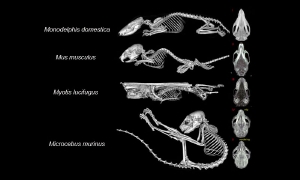 Skeletal views from the opossum, mouse, bat and lemur
He is corresponding author of a multi-institutional paper on the subject in the May issue of The Anatomical Record. The journal features a microCT image on the cover.
“This is a paper about how scientists scrutinize birth defects,” said Dr. Keller, assistant professor of cellular and structural biology at the UT Health Science Center’s Greehey Children’s Cancer Research Institute. “We measured differences in skull and limb shape and length between very different types of animals that are commonly studied by geneticists. These techniques, which we developed, serve as a set of standards for geneticists who wish to assess other important bones, such as the bone that is commonly malformed in cleft palate or other face and skull deformities.”
The genetics revolution is enabling developmental biologists and geneticists to more rapidly conduct a host of gene function experiments, such as changing the expression or activity of a gene to make a limb longer or shorter. By doing this, scientists hope to more precisely determine a gene’s function in both normal and abnormal physiology. “These studies will help us understand birth defects, how to prevent them and how to treat them,” Dr. Keller said.
The paper includes skeletal images of a mouse, a zebrafish, a chicken, a duck, the little brown bat, the African clawed frog, an opossum and the mouse lemur. The research team obtained some of the skeletal specimens from the University of Utah, the Southwest Foundation for Biomedical Research in San Antonio and the Lemur Center at Duke University.
Advanced image analysis techniques were made possible by collaboration with the Scientific Computing and Imaging Institute at the University of Utah.
“The microCT is excellent for high-resolution, morphological (structural) study of developing bones without the introduction of artifacts of decalcification and tissue processing,” said Frank J. Weaker, Ph.D., associate professor of cellular and structural biology at the UT Health Science Center San Antonio.
Dr. Weaker is an anatomy lecturer and course director whose own educational approach has been to teach dental and medical students using computed tomography-based three-dimensional images.
The paper includes the contributions of more than a dozen investigators from the UT Health Science Center San Antonio Greehey Children’s Cancer Research Institute, the Southwest Foundation for Biomedical Research, the University of Utah and George Washington University.
A laureate for the 2007 Nobel Prize in Physiology or Medicine, Mario R. Capecchi, Ph.D., of the Department of Human Genetics at the University of Utah, is a co-author. Dr. Keller joined the UT Health Science Center from Dr. Capecchi’s laboratory in January 2005.
The work was made possible in part by software from the National Institutes of Health National Center for Research Resources (NCRR) Center for Integrative Biomedical Computing.