Bright-field microscopy uses light to produce a dark image against a bright background. Often considered one of the simplest types of microscopy, a bright-field microscope uses an objective, condenser and eyepiece to magnify the image of a sample so the eye can see more minor features.
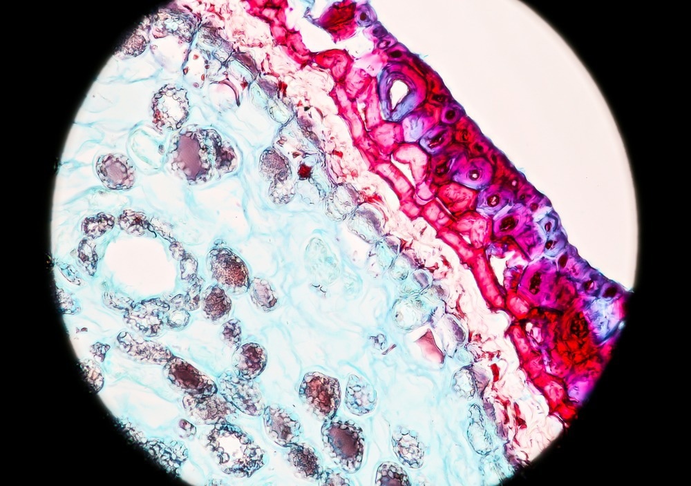
Pine stem cross-section microscopy bright field magnification 400X. Image Credit: Power_J/Shutterstock.com
A bright field microscope uses a transmission measurement to visualize the sample, where the light passes directly through. The contrast in the final image observed by the eye originates from different regions of the sample absorbing different amounts of the incident light.
While using transmission geometries in microscopy does limit the sample thickness that can be measured, one of the critical advantages of bright-field microscopy over other imaging methods such as fluorescence is that the sample preparation is very straightforward.
Before imaging, bright-field microscopy does not require sample staining or fluorophores to label particular cellular structures.
The simple-to-use, relatively compact, and affordable microscopes have made bright-field microscopy widely used in several fields, including food science1, soil science2, and medical applications.3
Principles of Bright-Field Microscopy
An optical bright-field microscope consists of a light source, a condenser, the sample stage and mount, objectives, and a mount.
The light source is pointed upwards from the bottom of the instrument and placed beneath the sample stage. Typical light sources for optical measurements include halogen lamps or light-emitting diodes (LEDs).
The condenser collects as much of the light from the source as possible and focuses this onto the sample.
When using highly divergent light sources such as lamps or LEDs that do not behave at point sources, it is necessary to use a condenser rather than a single lens to produce a well-focused beam. Specialist condensers can also help correct aberrations in the beam to improve the overall resolution of the measurement or to create specific beam shapes if necessary.
Once the light has passed through the sample, it enters the objective, which determines the ultimate magnification achieved with the microscope.
The objective and eyepiece magnify the sample image that the eye can view. There can be a trade-off between achieving a greater field of view of the sample and seeing more significant regions simultaneously as achieving maximum magnification.
The right choice of eyepiece can also help correct image distortions and be used for color correction and ensuring images appear sharp.
There is a tradeoff in the eyepiece cost and quality of the lenses used to achieve good performance. To make microscopy portable for point-of-care devices as well as more affordable, methods using smartphone cameras for both light sources and imagers have been developed.4 Such instruments can be used for live cell imaging and visualizing species such as zooplankton, making them a powerful tool for in field measurements.
Image Visualization
Some of the challenges of the method include blurry images of out-of-focus regions of the samples and weak absorbers can be challenging to image as they do not have sufficient contrast on the bright background. Molecules such as eosin and hemoglobin are much easier to image with optical bright-field approaches as they have large absorption cross-sections and produce a good background contrast even with very thin samples.5
Bright-field microscopy methods do not just have to be used with optical techniques but are also compatible with electron-based microscopy approaches, such as transmission electron microscopy. 6
Electron microscopes often have better spatial resolutions than are achievable with optical microscopes, as the diffraction limit of visible light is in the hundreds of nanometers. It is preferable to use electrons rather than optical sources for imaging nanoscale structures that have features less than 100 nm in size with sufficient resolution.
The set-up for bright field electron microscopy image visualization is similar to an optical microscope, only the light source is replaced with a high-power electron gun and images are recorded on an imaging plate or camera.
A set of electron lenses that serve a similar function to the condenser, objective and eyepiece are used for controlling the beam shape in the instrument and ensuring accurate image visualization of the sample is achieved with good resolution.
The simplicity and wide applicability of bright field imaging means it is likely to remain a workhorse technique in many different areas, including medical diagnostics.
The use of stains can help overcome some of the limitations associated with trying to image species that are weak absorbers and increase the number of specimens that bright field microscopy can be used for.
Two areas of bright field microscope development are finding ways of further automating the focusing and image analysis for higher sample throughputs and finding ways to make microscopes more compact, such as using smartphones.
References and Further Reading
- Shen, C., & Zhang, Y. (2017). Staining technology and bright-field microscope use. In Food Microbiology Laboratory for the Food Science Student (pp. 9-14). Springer, Cham. https://doi.org/10.1007/978-3-319-58371-6_2
- Stoops, G. (2021). Guidelines for analysis and description of soil and regolith thin sections (Vol. 184). John Wiley & Sons. https://doi.org/10.1002/9780891189763
- Farahani, N., Parwani, A. V., & Pantanowitz, L. (2015). Whole slide imaging in pathology: advantages, limitations, and emerging perspectives. Pathol Lab Med Int, 7(23-33), 4321. https://doi.org/10.2147/PLMI.S59826
- Orth, A., Wilson, E. R., Thompson, J. G., & Gibson, B. C. (2018). OPEN A dual-mode mobile phone microscope using the onboard camera flash and ambient light. Scientific Reports, 1–8. https://doi.org/10.1038/s41598-018-21543-2
- Hard, R., Hipp, J., Tangera, M., & Tomaszewski, J. (2014). Applications of Image Science in Pathology and Cell Biology. Elsevier Inc. https://doi.org/10.1016/B978-0-12-386456-7.07203-8
- Findlay, S. D., Shibata, N., Sawada, H., Okunishi, E., Kondo, Y., & Ikuhara, Y. (2010). Ultramicroscopy Dynamics of annular bright field imaging in scanning transmission electron microscopy. Ultramicroscopy, 110(7), 903–923. https://doi.org/10.1016/j.ultramic.2010.04.004
Disclaimer: The views expressed here are those of the author expressed in their private capacity and do not necessarily represent the views of AZoM.com Limited T/A AZoNetwork the owner and operator of this website. This disclaimer forms part of the Terms and conditions of use of this website.