Phase contrast microscopy manipulates phase shifts in light as it passes through transparent specimens. By converting phase shifts to changes in amplitude (brightness), the method enables scientists to view organic and biological samples with high levels of detail without needing sample preparation, staining, or labeling.
Image Credit: Catalin Rusnac/Shutterstock.com
How Does Phase Contrast Microscopy Work?
Dutch physicist Frits Zernike, awarded the Nobel Prize in Physics in 1953 for his work, invented the phase contrast microscope in the early 1930s.
Zernike noted that light waves traveling through a medium (not a vacuum) interact with the medium and experience wave amplitude and phase shifts depending on the properties of the medium.
Amplitude or brightness changes result from light being scattered and absorbed when it interacts with the medium. These changes often depend on the precise wavelength of light emitted and may result in different colors being seen.
Phase changes are not visible to the human eye or optical photographic equipment but can be viewed with phase contrast microscopy. These changes convey helpful information about the medium.
To make phase changes visible for study in phase contrast microscopy, a device is required to separate illuminating (background) light from the light scattered through interaction with the specimen (in the foreground).
A ring-shaped illuminating light is passed through a condenser annulus and focused onto the specimen. Some of this light is scattered by the specimen (foreground light), and the rest (background light) is unaffected.
The scattered light is weak and generally phase-shifted by −90° relative to background light. As a result, the foreground and background have a similar intensity, which results in low image contrast.
Contrast can be generated by limiting the amount of background light reaching the image plane, or by creating a constructive interference between background and foreground light.
It is also possible to achieve phase contrast microscopy results by phase shifting light by +90°.
Advantages of Phase Contrast Microscopy
A significant advantage of phase contrast microscopy is that any living cells can be studied naturally without any requirements for fixation, labeling, or staining.
All images generated by the technique can be label-free. This also saves time for researchers and reduces opportunities for mistakes or variations between experiments.
Phase contrast works well with transparent samples, as the additional interference makes the sample more visible. The entire object is illuminated in this technique.
For studying intracellular components of living cells, phase contrast microscopy offers relatively high levels of resolution. Cell mitochondria, mitotic chromosomes, vacuoles, and other living cell components can be studied with this method.
Components for phase contrast microscopy can also be added to nearly every type of optical microscope, as long as the phase objectives conform to the microscope’s tube length parameters and the condenser can be married to the correct-sized annular phase ring. This makes it a flexible and widely applicable technique.
Disadvantages of Phase Contrast Microscopy
Phase contrast microscopy is costly, with objectives running at $1,000.
There are also distortionary effects, such as a halo light ring that appears as a natural consequence of creating phase contrast. This distortion can be corrected using costly specialist equipment featuring an external interferometric module.
What is Phase Contrast Microscopy Used For?
Most of the applications for phase contrast microscopy are in biology research and education. Since its invention, the technique has enabled biologists to study living cells, watching them multiply through cell division and progress through life stages in real-time.
Below are some of the critical application areas for phase contrast microscopy.
Observing living cells
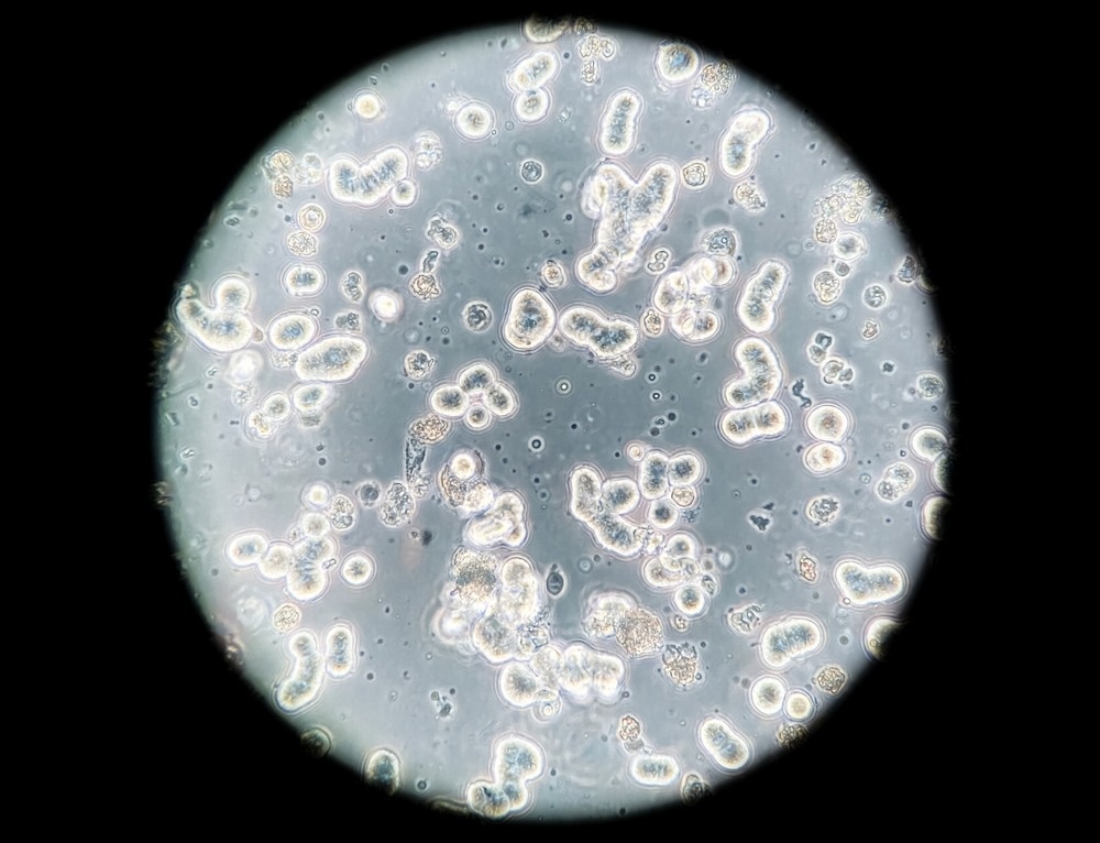
Human breast cancer cells cultured in vitro. Image Credit: Artur Wnorowski/Shutterstock.com
Phase contrast microscopy helps observe living cells in cultures and how they interact and grow within the culture.
Similarly, it is used for analyzing microorganisms’ cellular structures and to image latex dispersions in cells.
Before the introduction of phase contrast microscopy, biological cells could only be imaged with prior staining. As well as requiring additional preparation, this can lead to the death of many biological cells.
On the other hand, phase contrast can be used with living cells as staining is not required.
Engineers from Universidad de Guanajuato, Mexico, proposed an artificial intelligence (AI)-powered method for automatically identifying cells and tracking cell migration using phase contrast microscopy.
The team obtained data from several cells with phase-contrast video microscopy and introduced a method that classifies them based on variance. Once cells had been detected, their algorithm then tracked cell migration by finding their centroids.
Materials science
Phase contrast microscopy is also applied in materials science by visualizing surface structures with minimal sample preparation.
Materials scientists use the method when developing advanced or bionic fibers by analyzing and imitating natural fibers. Glass fragments are also good samples for this technique.
Paleontology
Paleontologists also use phase contrast microscopy to document fossils’ lithographic patterns.
References and Further Reading
Ambrez-Colin, F., et al. (2006). Detection of Biological Cells in Phase-Contrast Microscopy Images. Fifth Mexican International Conference on Artificial Intelligence. doi.org/10.1109/MICAI.2006.12.
Ingle, R. (2022). The Principles and Applications of Phase-Contrast Microscopy. [Online] AZO Optics. Available at: https://www.azooptics.com/Article.aspx?ArticleID=2123 (Accessed on 21 October 2022).
Introduction to Phase Contrast Microscopy. [Online] Nikon MicroscopyU. Available at: https://www.microscopyu.com/techniques/phase-contrast/introduction-to-phase-contrast-microscopy (Accessed on 21 October 2022).
Principle And Applications Of Phase Contrast Microscopy. [Online] SMACgig World. Available at: https://www.smacgigworld.com/blog/principle-and-applications-phase-contrast-microscopy.php (Accessed on 21 October 2022).
Ward, B. Phase Contrast Microscopy: A Simple Explanation. [Online] Microscope Clarity. Available at: https://microscopeclarity.com/phase-contrast-microscopy-a-simple-explanation/ (Accessed on 21 October 2022).
Zernike, F., (1955). How I Discovered Phase Contrast. Science. doi.org/10.1126/science.121.3141.345.
Disclaimer: The views expressed here are those of the author expressed in their private capacity and do not necessarily represent the views of AZoM.com Limited T/A AZoNetwork the owner and operator of this website. This disclaimer forms part of the Terms and conditions of use of this website.