The nutritional content of food is proportional to the number of nutrients stored in the food. Understanding this nutritional content is important for determining product labeling and ensuring quality control of produce. The nutritional content of cereals is particularly important as they provide a significant proportion of the world’s total calorie requirements and 50% of the world’s crop production.
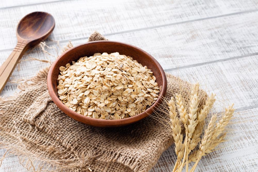
Image Credit: Vladislav Noseek/Shutterstock.com
There are several methods for determining the nutritional content and quality of cereal crops and many of these involve different microscopy techniques.2 Optical microscopy enables visual inspection of the grains and, with the right microscopy technique, identification of the amount of starch and other nutrients.
Food Microscopy
Food microscopy characterizes the properties and quality of food items, including contaminants or spoilage of food products. Food microscopy does not involve using a single microscopy technique, but a series of techniques, including scanning electron microscopy, transmission electron microscopy, fluorescence and optical microscopy.2
The reason such a wide range of techniques need to be used to determine the nutritional quality of cereals is that they provide complementary information.
Scanning electron and transmission electron microscopy provide much greater degrees of magnification of the sample than is possible with optical microscopy.
Such high levels of magnification and high spatial resolutions of sub-100 nm are necessary for full nutritional analysis cereal quality.3 The need for such high spatial resolution is due to the structural compounds that make up the main nutritional content of cereals – starches, fibers, fats, oils, etc. – are on this length scale.
Scanning electron microscopy is particularly well-suited to looking at food samples because it provides information on the surface structure and internal composition of the cereal. Microscopy methods can be used on both raw and processed cereals to identify if heating or freezing has caused damage to the nutritional structures.
Composition Analysis
While standard optical microscopy methods may not achieve the same spatial resolutions as electron-beam microscopy approaches, optical microscopy is invaluable in food nutrition analysis.
The sample preparation for optical microscopy typically involves taking a slice or cross-section of the cereal of interest.
Stains are then added to the sample to identify particular species of interest. Staining makes it possible to identify different types of proteins and the presence of compounds such as insulin and build up a nutritional profile of the sample as well as its distribution in the food.4 Optical confocal microscopy is used as a cost-effective way to acquire these images and is particularly valuable for looking at spatial distributions of nutrients and their dispersion through the sample.
Fluorescence microscopy is a commonly chosen technique to look for the presence of adulterants or contaminants in a food sample. Physical contaminants such as glass, stones or dirt can be identified with optical microscopy, but contamination with chemical species is much easier to identify with fluorescence. Many nutrients are also inherently fluorescent, such as many vitamin species.
The amount of fluorescence from a sample is proportional to the concentration of the fluorescing species; with the right calibration measurements, fluorescence microscopy can be used for quantitative and qualitative analysis of both nutritional and contaminant species.
In food samples where the nutritional species of interest does not fluoresce, fluorescence tags or labels can be used. These are highly fluorescent species, often chemical dyes, that bind to specific chemical species. Normally their fluorescence profile changes on binding to make it possible to distinguish between unbound, free tags and those attached to the species of interest.
Fluorescence microscopy can be used for sugar residues, bacteria and even physical properties of food samples such as emulsion stability.5
As well as static analysis of foodstuffs and their nutritional properties, microscopies are also being used in dynamical studies to look at how many of the properties of a food sample change when exposed to different environmental conditions such as structural deformations or phase changes.5
The Future of Food Microscopy
As there is no perfect microscopy method for looking at all food samples or creating a complete nutritional profile of a sample alone, there are still new microscopy variants being developed for food microscopy.
This includes methods such as confocal Raman microscopy, which can provide spectroscopic information, as well as imaging and atomic force microscopy.
There have been great efforts to breed crops with improved nutritional profiles, particularly for regions that are routinely affected by famines. Food microscopy is key to this work as it can be used to perform a quantitative analysis of the different levels of nutrients in samples as well as to detect what changes certain genetic modifications have had on the nutritional structures in the food.
References and Further Reading
- Mckevith, B. (2004). Nutritional aspects of cereals. Nutrition Bulletin, 29, 111–142. https://doi.org/10.1111/j.1467-3010.2004.00418.x
- Yiu, S. H. (1993). Food Structure Food Microscopy and the Nutritional Quality of Cereal Foods. Food Structure, 12, 123–133. https://digitalcommons.usu.edu/foodmicrostructure/vol12/iss1/13
- Sharma, V., & Bhardwaj, A. (2019). 29 - Scanning electron microscopy (SEM) in food quality evaluation. In Evaluation Technologies for Food Quality. Elsevier Inc. https://doi.org/10.1016/B978-0-12-814217-2.00029-9
- Guardeño, L. M., Vázquez-gutiérrez, J. L., & Hernando, I. (2013). Effect of Different Rice Starches, Inulin, and Soy Protein on Microstructural, Physical, and Sensory Properties of Low-Fat, Gluten, and Lactose Free White Sauces. Czech Journal of Food Science, 31(6), 575–580.
- Auty, M. A. E. (2013). Confocal microscopy: principles and applications to food microstructures. Food Microstructures. https://doi.org/10.1533/9780857098894.1.96
Disclaimer: The views expressed here are those of the author expressed in their private capacity and do not necessarily represent the views of AZoM.com Limited T/A AZoNetwork the owner and operator of this website. This disclaimer forms part of the Terms and conditions of use of this website.