Atomic force microscopy (AFM) is a powerful microscopy technique. It is used to acquire high-resolution images at the nanoscale to better understand the properties of matter.
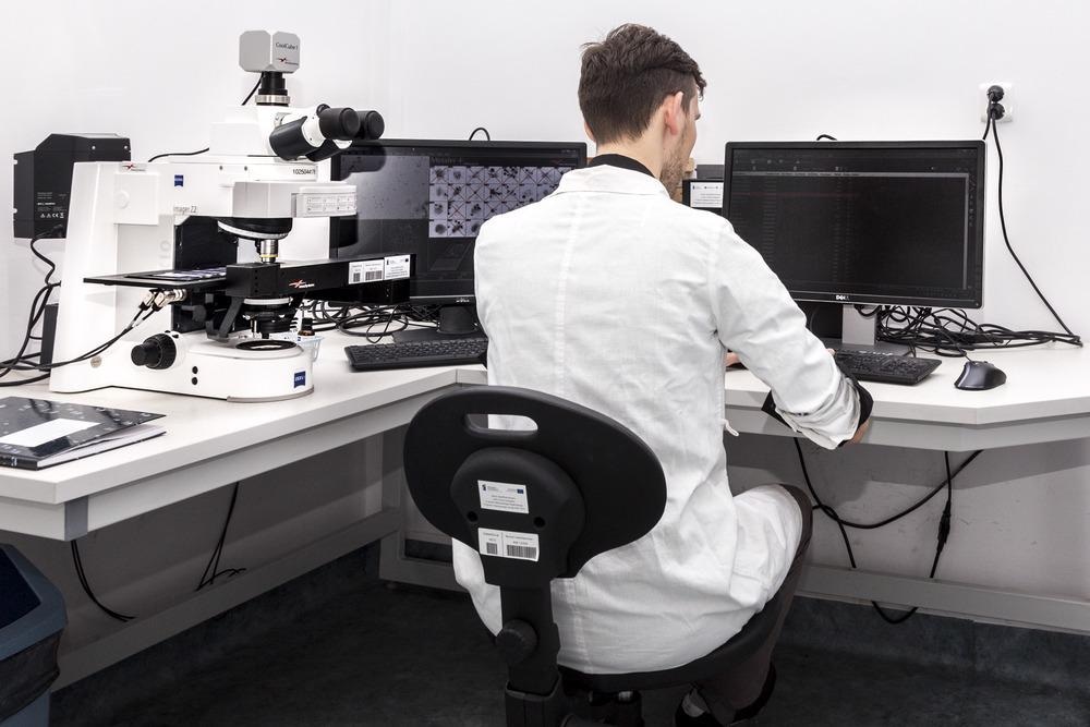
Image Credit: dominika zara/Shutterstock.com
AFM was first introduced in a breakthrough experiment in 1986. Gerd Binnig and Heinrich Rohrer from IMB-Research had earlier garnered a Nobel prize for their invention of Scanning Tunnelling Microscope (STM). However, the STM could only image metal or semiconductor materials. Therefore, Binnig, along with Christoph Gerber and Calvin Quate developed the AFM. The AFM builds on the capabilities of the STM but can be used to image any very high-resolution material. AFM images can resolve an image a million times smaller than a human hair.
Applications of Atomic Force Microscopy
Many scientific processes can be improved when the underlying mechanisms are understood better. With better knowledge of the material structure, new and useful technology and devices can be developed.
AFM is widely used for imaging friction between surfaces, the viscosity of liquids, the elasticity of materials, electrical forces, magnetic forces, conductivity, capacitance, and surface potential, among many other properties.
Applications of AFM include imaging:
- Biological molecules
- Cellular components
- Cells or tissues in biochemistry applications
- Polymers
- Nanostructures
- Other materials in chemistry, materials science, nanotechnology, and cancer research
Set-up and Working Principles of Atomic Force Microscopy
An AFM consists of four key components, the first being a cantilever with a sharp tip. The back of the cantilever is coated with a reflective material so that it reflects light like a mirror. A laser is directed to the back of the cantilever and the reflected light is then collected by a photo-detector, which is very sensitive to the positions of the laser beam. The cantilever, laser, and detector system are precisely scanned by an electric controller. The sample, sitting under the microscope on a translation stage, can also be moved.
Figure 1 illustrates a typical AFM setup. The cantilever is used as a force sensor. When the tip of the cantilever is scanned across a sample, the tip acts like an elastic.
Depending on the amount of force between the tip and the sample, the cantilever compresses and stretches. The photo-detector records the changes to the reflected laser beam position proportional to the movement of the cantilever. A detailed topographical image of the sample can be captured by scanning across the surface of the material.
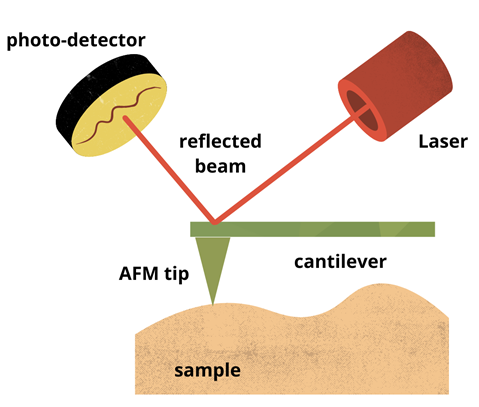
Figure 1: Schematic illustrations showing basic principles of AFM. Image Credit: Ilamaran Sivarajah
Type of Forces Detected
Different types of forces can be detected between the AFM tip and the sample, including electrostatic forces between charges on the surface of the sample and the tip. Other forces detected include van der Waals forces, mechanical contact force, capillary forces, magnetic forces, Casimir forces, and forces induced by chemical bonds. The AFM can be operated in different modes to best suit the type of force that is measured.
Operating Modes of AFM
Static mode
In static mode, also called contact mode, the AFM tip is in contact with the sample surface when it is scanned. The tip is “dragged” across the sample like a needle. The tip acts like an elastic, compressing and stretching when it encounters a force induced by the sample surface. The changes in the cantilever deflect the laser beam which is imaged on the photo-detector.
Soft materials are deflected further as force is applied, so AFM tips are generally made from soft materials. But since the tip is in contact with the sample it is scanned across, the sample can be damaged easily. The static mode is mostly used to image hard samples to obtain topographical information.
Dynamic mode
In dynamic mode, the cantilever is driven to oscillate at a certain frequency instead of being “static”. Within the dynamic mode of operation, tapping mode and non-contact mode are widely employed.
In tapping mode, the tip oscillates up and down above the surface of the sample. The tip comes in contact with the sample at its lowest point of oscillation by “tapping” the surface. Repulsive forces are detected when the tip “taps” the surface, while attractive forces are detected at the peak of oscillation. Tapping mode generally lessens the damage done to a surface and the tip compared to contact mode. This technique is used to image the formation of molecules in chemistry among many other applications.
In non-contact mode, the tip of the cantilever does not contact the sample surface. The cantilever is driven to oscillate above the surface of the sample. Therefore, the tip does not cause any damage to the sample. In non-contact mode, the driving motion of the cantilever is either amplitude-modulated or frequency-modulated.
These modulation techniques offer different detection sensitivities dependent on the application. The non-contact measurement method is advantageous for imaging soft samples. Some applications include imaging of biological samples and organic thin film.
The operational modes of the AFM are illustrated in Figure 2.
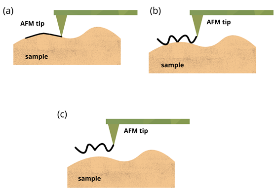
Figure 2: Schematic illustrations showing the operational modes of AFM (a) Contact mode, (b) Tapping mode, (c) Non-contact mode. Image Credit: Ilamaran Sivarajah
Challenges and New Developments of Atomic Force Microscopes
Along with causing sample damage, the AFM has other drawbacks. The size of an AFM image that can be captured in a single scan is small compared to other scanning microscopy methods.
AFM scan speeds are also limited because of the mechanical integration of the cantilever and the detection apparatus. Due to the oscillation of the tip, hysteresis can also affect the performance of the AFM.
AFM can be combined with a variety of experimental techniques developed in other scientific efforts, including optical microscopy and spectroscopy techniques such as fluorescent microscopy or infrared spectroscopy. These combined methods have resulted in new explorations such as scanning near-field optical microscopy.
Adapting single-photon excitation methods adapted from condensed matter physics has further expanded the applicability of AFM.
Non-optical detection methods such as qPlus-based AFM with functionalized tips have also been successfully implemented.
Advancements in AFMs promise to advance investigations of photovoltaics and energy-storage research, polymer sciences, nanotechnology, and medical research.
References and Further Reading
Jinbo Peng, Jing Guo, Runze Ma, Ying Jiang (2021) Water-solid interfaces probed by high-resolution atomic force microscopy. Surface Science Reports, 100549, ISSN 0167-5729. https://doi.org/10.1016/j.surfrep.2021.100549
Liao, M., Nicolini, P., Du, L. et al. (2021) UItra-low friction and edge-pinning effect in large-lattice-mismatch van der Waals heterostructures. Nat. Mater. https://doi.org/10.1038/s41563-021-01058-4
McClelland G.M., Erlandsson R., Chiang S. (1987) Atomic Force Microscopy: General Principles and a New Implementation. In: Thompson D.O., Chimenti D.E. (eds) Review of Progress in Quantitative Nondestructive Evaluation, vol 6 A. Springer, Boston, MA. https://link.springer.com/chapter/10.1007/978-1-4613-1893-4_148
Takeshi Fukuma (2010) Water distribution at solid/liquid interfaces visualized by frequency modulation atomic force microscopy, Science and Technology of Advanced Materials, 11:3. https://doi.org/10.1088/1468-6996/11/3/033003
Disclaimer: The views expressed here are those of the author expressed in their private capacity and do not necessarily represent the views of AZoM.com Limited T/A AZoNetwork the owner and operator of this website. This disclaimer forms part of the Terms and conditions of use of this website.