Article updated on 17 August 2021
Mass spectrometry is a key workhorse technique in analytical sciences. Waters has released a multi-reflecting time-of-flight (MRT) mass spectrometry platform to deliver improved resolution with short acquisition speeds.1
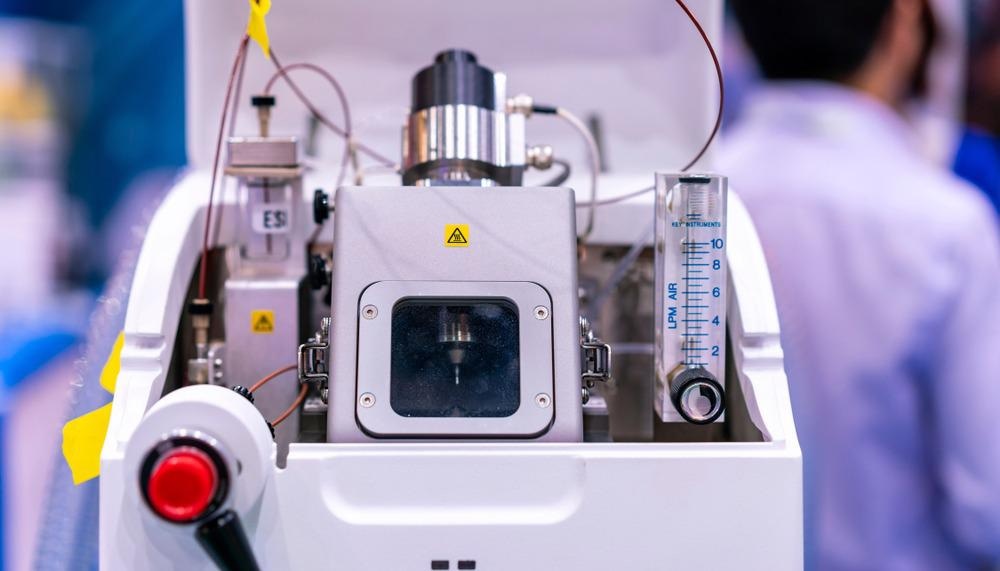
Image Credit: Surasak_Photo/Shutterstock.com
High-Resolution Mass Spectrometry
A mass spectrometer with sufficiently high resolution can measure masses significantly closer to the ‘actual mass’ of a compound than a lower resolution instrument. The closer a measurement is to the actual mass, the greater the discriminating power of the mass spectrum to differentiate between even very similar chemical species. For example, many chemical compounds have identical nominal masses, but their actual mass-to-charge ratios (m/z) may differ by 0.001. This difference can be detected using a sufficiently high-resolution instrument.
The new Waters Select Series MRT offers parts per billion mass accuracy to maximize confidence in sample identification. The Select Series MRT achieves ultra-high-resolution measurements without requiring longer scan times. Measurements can be made at a 10 Hz sampling rate without compromising the 200,000 Full Width Half Maximum resolution that is entirely independent of the scan speed.2
Sample Delivery
Mass spectrometry is typically performed on gaseous ions. Therefore, for most biological samples that are typically solids or liquids, the sample needs to be ionized for the measurement process. There are several methods to this for mass spectrometry that vary in the degree of fragmentation they cause to the ion. Harder ionization techniques lead to a greater degree of smaller fragment ions, which can be helpful for the chemical identification of some compounds, but larger molecules can produce highly congested images.
One technique that is very popular for recording mass spectra of biological compounds is Desorption Electrospray Ionization (DESI). One of the key advantages of this technique is that it can be used under ambient conditions. DESI works by firing a fast-moving charged solvent stream at the sample of interest to extract analytes from the surface that can be transferred for mass analysis.
It is possible to also extend DESI to work in an imaging mode. This is done by placing the sample of interest onto a translation stage and selectively ionizing certain localized regions. The sample can then be rastered until a full image of its whole area has been recorded. Mass spectrometry imaging techniques such as DESI are proving powerful emerging tools for the characterization of biological specimens.
Another ionization technique that is compatible with both imaging and spectrometry modes is Matrix-Assisted Laser Desorption Ionization (MALDI). Rather than using a charged stream of solvent to extract and ionize analytes, MALDI uses a laser source to desorb analytes from a surface.
First, the analytes of interest are prepared in a MALDI-compatible matrix. When this is irradiated with a laser beam, the matrix absorbs the light, converting it to heat energy that is then transferred to the analyte to vaporize it. Like DESI, MALDI can also be used under ambient conditions. Preparation of the sample matrix means that a small amount of additional sample preparation is required but these procedures can be relatively straightforward and quick to do.
MALDI has become widely used in clinical microbiology labs as a high-throughput, low-cost measurement technique as it can be used to identify and differentiate between many strains of bacteria. It also shows promise for other types of pathogens, including viruses and fungi.
Waters Advantage
The Waters Select Series MRT comes with both DESI and MALDI imaging sources for maximum flexibility when studying tissue samples. When combined with the extreme high-resolution mass spectrometer, this combination allows for unparalleled clarity for tissue imaging, making it possible to investigate complex processes such as how targeted therapeutics operate.
The DESI XS imaging source has been optimized for highly efficient analyte transfer to give excellent sensitivity and for compatibility with a wide range of chemical surfaces. DESI is particularly well-suited to the study of small biological molecules such as lipids and metabolites and can be used to visualize the spatial distribution of these within tissue samples. As no additional sample preparation is required, this makes the DESI XS source an excellent candidate for high throughput measurements.
The Walters MALDI imaging source can help visualize relatively large 10-micron tissue sections and is better suited for the measurement of high molecular weight species, such as peptides and proteins. By using MALDI and DESI imaging in tandem, it is possible to generate highly informative images that capture both the local chemical and structural information of the tissue samples of interest.
The combination of the DESI and MALDI imaging sources with this quality of detection makes the Waters Select Series MRT the first commercial instrument available of this type. This new instrument draws on Waters’ extensive expertise in specialized measurements and mass spectrometry for the life sciences to offer new opportunities for research in this area by making high-resolution mass spectrometry more accessible for the biological sciences.
References and Further Reading
- Waters (2021) Select Series MRT, https://www.waters.com/waters/en_GB/SELECT-SERIES-MRT/nav.htm?cid=135082877, accessed 18 June 2021
- Waters (2021) Waters Sets New Standards, https://www.businesswire.com/news/home/20210608006036/en/, accessed 18 June 2021
Disclaimer: The views expressed here are those of the author expressed in their private capacity and do not necessarily represent the views of AZoM.com Limited T/A AZoNetwork the owner and operator of this website. This disclaimer forms part of the Terms and conditions of use of this website.