Image Credit: Okrasiuk/Shutterstock.com
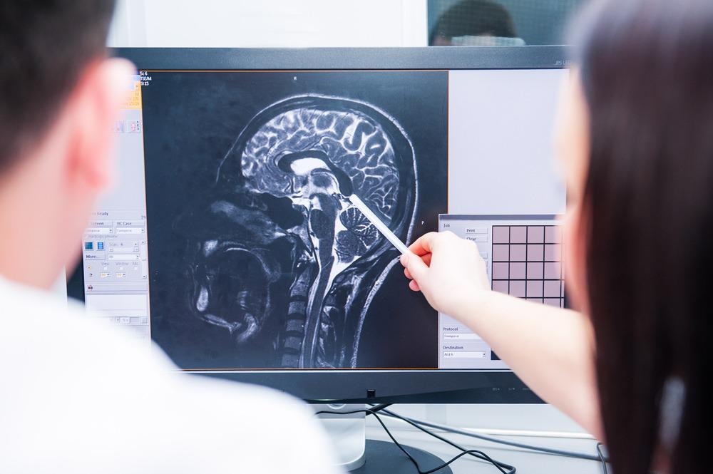
Scientists at the University of California - Davis have developed an innovative, non-invasive method of assessing brain injuries using light. The new system measures brain blood flow without the need for invasive surgery and is cheaper than existing non-invasive technologies. For this reason, the revolutionary new technique developed by biomedical engineers and neurologists will likely have a significant impact both in the field of neuroscience research and in the area of medical diagnostics, disease monitoring, and particularly, in the assessment of brain injuries.
The “Silent Epidemic” of Traumatic Brain Injuries
Each year, roughly 2.5 million people acquire a traumatic brain injury (TBI), which is the leading cause of death and disability in those aged from 1 to 44 years. It is also a major cause of death, with around 50,000 people dying each year due to TBI and a further 80,000 suffering permanent disability.
According to the Centers for Disease Control and Prevention (CDC), the acquirement of a moderate to severe TBI can often lead to long-term or life-long health effects and care is generally needed to facilitate recovery. Overall, TBI carries huge health and economic burdens.
Considered a “silent epidemic”, TBI is the world’s leading cause of death and disability in terms of traumatic injuries. However, much is still unknown about the nature of TBI. Scientists do not know the full incidence or distribution of TBI, nor do they fully understand its health implications, particularly its long-term effects.
A growing body of research is revealing the link between TBI and psychological illness, however, more research is needed to gain a comprehensive understanding of this relationship. While methods currently exist to assess brain injuries in a non-invasive way, they are often costly and have drawbacks in accuracy. Now, a new method has been established using light that promises to overcome such limitations.
Developing a New, Non-Invasive Method of Brain Imaging
A team of biomedical engineers and neurologists at the University of California, Davis, has established a technique of detecting brain activation with light. The revolutionary method, known as functional interferometric diffusing wave spectroscopy (fiDWS), will be more cost-effective than currently existing non-invasive alternatives.
The technique visualizes how the brain regulates blood flow using principles similar to those of functional magnetic resonance imaging (fMRI). Blood flow is vital to the functioning of the human brain, which is illustrated by the fact that, while the human brain only contributes to 2% of our body weight, it requires 15-20% of our blood flow.
For many decades, measurements of cerebral blood flow have been vital to diagnosing strokes and making predictions of secondary damage in cases of TBI and subarachnoid hemorrhages. These methods have become utilized in the monitoring of recovery from such events.
Current technology is limited in that it is expensive and that it is not suitable for continuous bedside monitoring. Alternative light-based technologies also have limitations in accuracy.
The new fiDWS method uses near-infrared light, which can penetrate the body’s tissues. By shining this type of light on the human forehead, the light becomes scattered by the tissue. This scattering is also caused by the blood cells flowing through the brain. The light that returns out of the brain and through the skull and scalp is then measured and analyzed to determine the blood flow in different areas of the brain.
This method alone produces a weak signal. So, to overcome this, the UC - Davis researchers combined it with interferometry, a measurement based on the phenomenon of the interference of waves. Light waves can superimpose, reinforce, and cancel each other out. In particular, a strong light wave can enhance a weak one and, therefore, boost its detected energy.
To achieve this method of increased sensitivity, the team split a laser beam into paths of "sample" and “reference”, the sample being the one that entered the patient’s brain, and the reference being the one that is routed to reconnect with the sample beam prior to entering the detector. The stronger reference beam, via interferometry, enhances the weak signal.
In a paper published this year in the journal Science Advances, the team demonstrated how its method allowed it to measure the output with a simple light-detection, the same type used in digital cameras, rather than expensive photon-counting detectors used in alternative methods. Software was then used to calculate the blood flow index within different cerebral locations.
Tests showed that the new technology can measure cerebral blood flow more rapidly and at deeper surfaces than currently available light-based alternatives. The technology successfully picked up on pulsating cerebral blood flow and identified changes in blow flow when patients were administered a mild increase in carbon dioxide. Finally, the method proved it could detect when participants were undergoing cognitive tasks, such as solving math problems.
Overall, the evidence is promising. The team at UC - Davis has established a new, non-invasive light-based technology for assessing cerebral blood flow that may prove to be fundamental in assessing brain injuries. Given that it promises to be cheaper and more accurate than currently available alternatives, this new technology may make a significant impact in medical and research fields.
References and Further Reading
Centers for Disease Control and Prevention. (2014). Report to Congress on Traumatic Brain Injury in the United States: Epidemiology and Rehabilitation. National Center for Injury Prevention and Control; Division of Unintentional Injury Prevention. Atlanta, GA. https://www.cdc.gov/traumaticbraininjury/pdf/tbi_report_to_congress_epi_and_rehab-a.pdf
Dewan, M., Rattani, A., Gupta, S., Baticulon, R., Hung, Y., Punchak, M., Agrawal, A., Adeleye, A., Shrime, M., Rubiano, A., Rosenfeld, J. and Park, K., 2019. Estimating the global incidence of traumatic brain injury. Journal of Neurosurgery, 130(4), pp.1080-1097. https://thejns.org/view/journals/j-neurosurg/130/4/article-p1080.xml
Measuring Brain Blood Flow and Activity With Light. Andy Fell. UC Davis. Available at: https://www.ucdavis.edu/news/measuring-brain-blood-flow-and-activity-light
Schiller JS, Lucas JW, Ward BW, Peregoy JA. Summary health statistics for U.S. adults: National Health Interview Survey, 2010. National Center for Health Statistics. Vital Health Stat. 2012;10(252). https://pubmed.ncbi.nlm.nih.gov/22834228/
Walz, R., 2008. Psychiatric disorders and traumatic brain injury. Neuropsychiatric Disease and Treatment, p.797. https://www.ncbi.nlm.nih.gov/pmc/articles/PMC2536546/
Zhou, W., Kholiqov, O., Zhu, J., Zhao, M., Zimmermann, L., Martin, R., Lyeth, B. and Srinivasan, V., 2021. Functional interferometric diffusing wave spectroscopy of the human brain. Science Advances, 7(20), p.eabe0150. https://advances.sciencemag.org/content/7/20/eabe0150
Disclaimer: The views expressed here are those of the author expressed in their private capacity and do not necessarily represent the views of AZoM.com Limited T/A AZoNetwork the owner and operator of this website. This disclaimer forms part of the Terms and conditions of use of this website.