Image Credit: Elizaveta Galitckaia/Shutterstock.com
Atomic force microscopy (AFM) is an analytical technique capable of analyzing surfaces down to the molecular level, and for years it has been effectively used to describe collagen found in the anterior cruciate ligament (ACL).
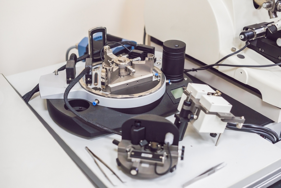
How Does AFM work?
AFM is frequently used to describe surface topography by passing an extremely fine probe tip connected to a cantilever over the top of a sample. Measuring of the cantilever deflection caused by the surface allows for the creation of a three-dimensional topographic image.
AFM produces nanoscale topological data that other analytical techniques cannot. For example, AFM can characterize the mechanical qualities of specimens at a nanoscale level, providing both qualitative and quantitative data. Moreover, AFM does not require specialized care of samples, such as labelling with fluorescent probes or the application of a special coating.
Analyzing ACL Collagen
Able to be conducted without causing damage to collagen, an AFM analysis can be used to investigate a broad array of collagen structures, from composite molecules to individual fibrils to collagen nanomaterials.
Used for different purposes, the most popular AFM techniques are contact mode (in which the probe tip is constantly contacting the sample), tapping mode (intermittent contact) and the noncontact mode. Tapping mode is more commonly used for soft biological specimens, as it is potentially less destructive than contact mode and less problematic than noncontact mode. Furthermore, AFM can evaluate hardness by pushing the tip down into the sample at a specific point.
With these abilities to gauge the hardness, elasticity and topography of specimens, AFM has become a popular tool in clinical research studies on collagen and other connective tissues. With collagen in particular, AFM can be used to determine the diameters, organization and overall morphology of collagen fiber bundles in three dimensions.
While electron microscopy has been used for the characterization of ACL collagen, as it can resolve topography almost as well as AFM, AFM has more advantages, including simpler sample preparation and the capability for nanomechanical assessment. Also, AFM imaging can offer clear imagery of collagen structures while avoiding lateral pressure on the specimens, which is particularly useful for the study of soft tissues.
As with many other analytical techniques, tissue sectioning is necessary for AFM testing. To get a clear AFM image, it is important to follow the fundamental procedures for tissue sectioning and fixation. Standard sample preparation for optical microscopy often translates to AFM. With comprehensive capabilities, particularly at the fibril level, AFM can dramatically depict unique features of ACL collagen.
The ability to produce 3D imagery is a particularly useful benefit for the AFM study of collagen structures. For example, two-dimensional images can display overlapping collagen fibrils, but not show them actually crossing in space. This is a distinctive feature of ACL grafts and the 3D reconstruction of these structures by AFM is crucial.
Using AFM to Study ACL Ruptures Caused by Repetitive Injuries
There is a common perception that ACL failures happen as a result of a singular, abrupt or dynamic movement, possibly influenced by insufficient neuromuscular training. However, research has shown serial ACL injuries create a 'ticking time bomb' situation, as consecutive minor injuries cause significant degeneration in the ligament, according to a July 2019 study in the American Journal of Sports Medicine.
The study team used AFM to examine ACL collagen in cadaver knees at the nanometer scale. According to their report, the study team could detect a consistent degradation at the molecular level of ACL collagen in knees that had successive injuries.
AFM found the dysfunction of collagen fibrils in the forms of decreased topographical surface solidity and the creation of sub-100-nanometer gaps in the collagen matrix of tested specimens.
The study team noted that AFM was particularly well-suited to their work and their conclusions could not have been reached without the technique.
References and Further Reading
Disclaimer: The views expressed here are those of the author expressed in their private capacity and do not necessarily represent the views of AZoM.com Limited T/A AZoNetwork the owner and operator of this website. This disclaimer forms part of the Terms and conditions of use of this website.