Article updated on 5 November 2020.

Shidlovski / Shutterstock
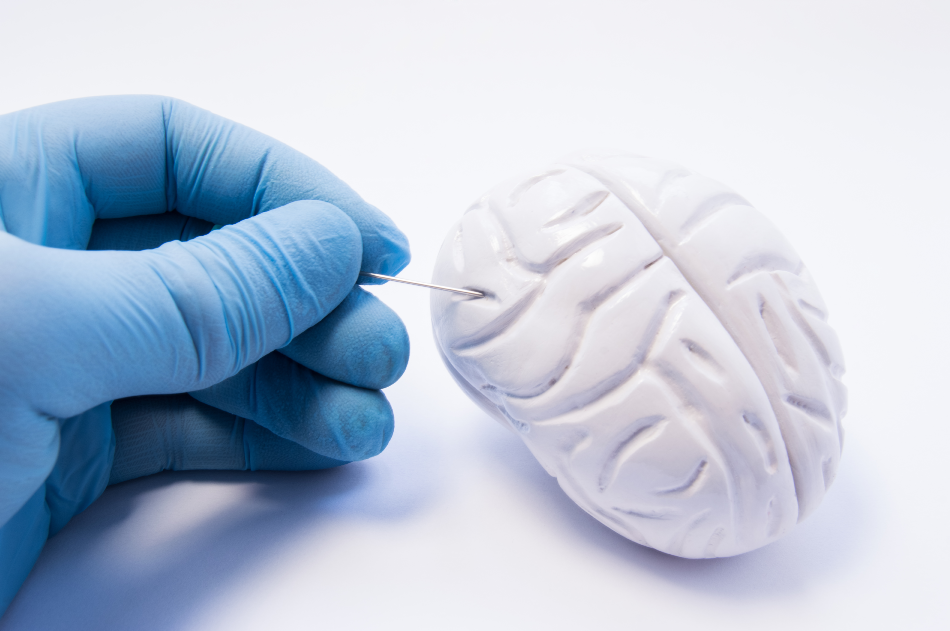
A Canadian team has developed a breakthrough in brain biopsy techniques with their Raman spectroscopy probe that enables surgeons to inspect brain tissue at the tip of the biopsy needle before they remove it.
An Innovation in Brain Biopsies
A new development in cancer treatment has emerged out of the work of a team of researchers from the Montreal Neurological Institute and Hospital alongside researchers at the Polytechnique Montreal. The Canadian research team has created a new system to enable neurosurgeons to determine malignant tissue in real-time before it is removed for stereotactic biopsy sampling. To achieve this, the team has developed a method that integrates a Raman spectroscopy guidance system within the tip of a brain biopsy needle.
The significance of what the team has achieved could potentially be huge for the treatment of brain tumors. The innovation would allow surgeons to collect biopsy samples from areas of the brain where the densities of cancerous cells are sufficient enough to achieve a reliable diagnosis while reducing the disruption caused to the surgical workflow. It is predicted that with this new technology, treatment plans could be optimized, and diagnosis through biopsies could prove to be more reliable.
Raman Spectroscopy in Cancer Treatment
In 1928 Dr. C. V. Raman published his discovery of a light scattering effect, known as the Raman Effect. He discovered that when a beam of light is deflected by molecules, a change in the wavelength of light occurs. The basic principle is that light passing through a transparent chemical compound sample is deflected in directions that are different from that of the incident. While most of the scattered light is of the same wavelength, a small portion presents wavelengths that are different from the incident light. This is because sometimes, the molecule takes or gives energy to the interacting photons, resulting in a small portion of the scattered light being of a higher or lower frequency. These changes in frequency can be related to the kind of sample the light has hit through Raman spectroscopy, providing a structural fingerprint of the molecule.
Uninterrupted Surgical Workflow and High-Quality Data
Raman spectroscopy is already used in the treatment of cancer as the identification of the molecule’s structural fingerprint can indicate the presence of gliomas or metastases. So far, the technique has been used to detect cancer of the colon, lung, and skin. Due to the nature of the technique which measures light scattering, Raman spectroscopy performs analysis in a non-destructive way, which makes it preferable for sampling living tissue.
The team in Montreal took this technology and successfully incorporated the tiny Raman probe (of about 900 μm in diameter) into the biopsy needle’s internal cannula. The needle they created is able to be guided by a navigation attachment which positions the needle using previously acquired MR images as a reference. Negative pressure can be created inside the needle through a Y-shaped sealed plastic breakout, enabling the surgeon to collect a sample of tissue without the need to retract the probe.
Validation that the new guidance system is effective has been provided by the Montreal team. A patient with glioblastoma and two patients with lymphoma have undergone surgical biopsy procedures using the device. The result was high-quality spectral data collected efficiently by the system.
Future Developments
An accurate statistical classification model is planned to be constructed in order to support the prediction of tissue type during brain biopsy in real-time. The system is expected to come onto the market very shortly, with Montreal’s ODS Medical set to commercialize it. Further future aims of the team include creating versions of the system to use with lung and prostate cancers.
Source
Disclaimer: The views expressed here are those of the author expressed in their private capacity and do not necessarily represent the views of AZoM.com Limited T/A AZoNetwork the owner and operator of this website. This disclaimer forms part of the Terms and conditions of use of this website.