A newly developed compact ISM microscope equipped with a single photon avalanche diode (SPAD) array detector provides high-resolution structural and functional imaging. Image Credit: A. Zunino (ITT)
The development of fast and compact detector arrays has sped up the field of laser scanning microscopy. By substituting the conventional single-element detector of confocal laser scanning microscopes with these arrays, new and distinct capabilities are made possible.
A pixelated detector records the spatial distribution of incident light in addition to the intensity value of the collected light, essentially creating a miniature image of the illuminated area at each scan point. Conventional detectors only provide the intensity value of the collected light.
The detector arrays give rise to additional spatial information that facilitates image scanning microscopy (ISM), a super-resolution method. ISM uses a raw multidimensional dataset that is generated by a microscope to computationally create a single image. Better optical sectioning, spatial resolution, and signal-to-noise ratio (SNR) are achieved in the final image construction than with a conventional confocal microscope. More specifically, the ISM image's lateral resolution can exceed Abbe's limit by a factor of two.
These benefits, however, are only realized by utilizing spatial information; time-resolved acquisition, which provides access to structural and functional information encoded into the fluorescence dynamics (such as fluorescence lifetime), can further enhance contemporary fluorescence bioimaging.
A new compact and efficient ISM microscope with a single photon avalanche diode (SPAD) array detector that can provide high-resolution structural and functional imaging in a single architecture was created by researchers at the Italian Institute of Technology (IIT) in Genoa. Advanced Photonics, a Gold Open Access journal, published the research.
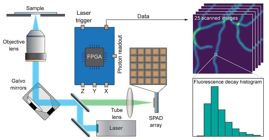
The described SPAD array detector is made up of a square grid made up of 25 separate diodes. The asynchronous read-out and small size allow for quick identification of fluorescence photons that are impinging. The data acquisition scheme is a heterodyne sampling technique based on the digital frequency domain (DFD) method. It allows the construction of the fluorescence decay histogram with a timing resolution of 400 ps, which is suitable for most fluorescence imaging applications.
The method is straightforward enough to enable the histogram to be computed on the same field-programmable-gate-array (FPGA) board that records the detected signal and operates the microscope, thereby streamlining the architecture of the device.
By combining ISM and time-resolved measurements with stimulated emission depletion (STED) microscopy using the separation by lifetime tuning (SPLIT) technique, the report highlights the versatility of the microscope. Without changing the acquisition plan, the outcome is an image with improved lateral resolution and contrast. Furthermore, multispecies imaging with a single detector for enhanced structural specificity is made possible by time-resolved measurements.
The phasor representation of fluorescence dynamics allows the system to distinguish between different dyes based on their fluorescence lifetime values. By using the pulsed-interleaving excitation technique, it is possible to distinguish between different fluorophores even when using dyes with similar lifetime values and overlapping excitation spectra. In fact, the spectral information is efficiently encoded into the temporal dimension by alternating laser excitation pulses of different colors. The two fluorescent dyes’ contributions can be later divided to prevent crosstalk because of the suggested microscope's exceptional time resolution.
The results of this work suggest that the future of laser scanning microscopy is tightly connected to SPAD array detectors, capable of enriching the microscopy dataset with additional spatial and temporal information without the need to change the optical architecture of a confocal microscope.
Giuseppe Vicidomini, Principal Investigator and Corresponding Author, Molecular Microscopy and Spectroscopy Lab, Italian Institute of Technology
The work demonstrates that SPAD array detectors combined with a tailored acquisition system make photon-resolved ISM easily accessible and usable.
Journal Reference
Tortarolo, G., et al. (2024) Compact and effective photon-resolved image scanning microscope. Advanced Photonics.doi.org/10.1117/1.AP.6.1.016003.