Medical imaging technology such as computed tomography (CT), magnetic resonance imaging (MRI), and X-Ray cannot offer real-time images during tumor resection surgery, so surgeons usually depend on touch, sight, and experience to recognize tumor margins, resulting in a high probability of tumor residues.
Schematic illustration of the Research. Image Credit: Prof. ZHANG’s group
In comparison with conventional medical imaging, near-infrared (NIR) fluorescence imaging has the benefits of real-time, high resolution, high sensitivity, no radiation, etc. Nevertheless, for tiny tumor residues, it needs relatively high sensitivity.
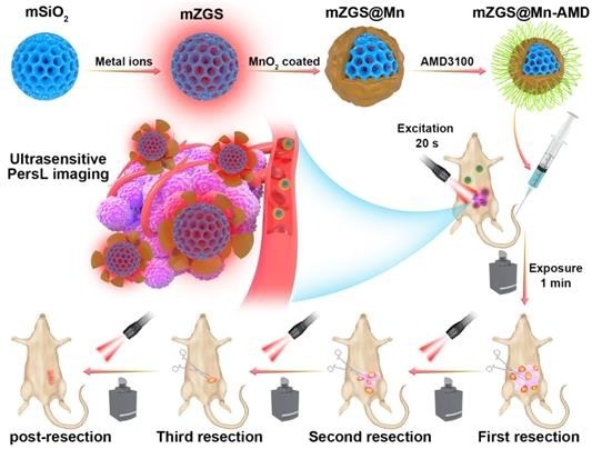
The study team headed by Prof. Yun Zhang from the Fujian Institute of Research on the Structure of Matter of the Chinese Academy of Sciences built a novel ultrasensitive nanoprobe (mZGS@Mn-AMD) with an ultrahigh tumor-normal tissue (T/NT) signal ratio to categorize residual tumor tissues from normal tissues. The research was published in Advanced Science.
Persistent luminescent nano core (mZGS) has high sensitivity as they do not require continuous excitation, and the background fluorescence interference produced by biological tissues is absent at the time of imaging.
The scientists quenched persistent luminescence (PersL) in normal tissue by the outer layer of MnO2 and recovered it owing to the degradation of MnO2 in the tumor microenvironment, enhancing the tumor imaging sensitivity.
The nanoprobe’s intraoperative tumor-to-normal tissue (T/NT) signal ratio was 58.8 in the orthotopic breast cancer model, which is around nine times that of down-conversion nanoparticles. The T/NT ratio of residual tumor (<2 mm) stayed at 12.4, significantly high to distinguish tumor tissue from normal tissue.
In addition, 4T1 liver-implanted tumor, multiple-microtumor, and lung metastasis models were made to establish that this ultrasensitive nanoprobe can identify tumor residues.
This research introduces convenient and ultrasensitive imaging for identifying residual tumor tissue, paving the path for entire surgical removal.
Journal Reference
Lin, P., et al. (2023) Near-Infrared Persistent Luminescence Nanoprobe for Ultrasensitive Image-Guided Tumor Resection. Advanced Science. doi.org/10.1002/advs.202207486.