The observation of living cells with the help of microscopes forms the basis of modern biology. Recent developments in optical microscopy enable cellular and sub-cellular imaging within model organisms like zebrafish, the vinegar fly or mice.
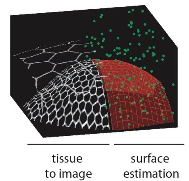 Left: Drawing of a curved biological tissue. The hexagons represent the fluorescent contours of the cells organized in a cell sheet. The tissue can be covered by a second epithelium which can be ignored by the imaging process. Right: From a few acquisitions (green dots), the microscope automatically estimates the surface of the tissue (red mesh) and can then concentrate the acquisitions on this surface, or even only on the fluorescent cell contours thanks to a propagative acquisition algorithm. Image Credit: Faris Abouakil, Huicheng Meng, Marie-Anne Burcklen, Hervé Rigneault, Frédéric Galland, and Loïc LeGoff.
The toxicity related to illumination is one of the main limitations of existing techniques, compromising the biological processes analyzed. Until now, there has been no solution to this problem except for decreasing the light level, which results in the loss of image quality.
A group of researchers headed by Dr. Loïc Le Goff and Dr. Frédéric Galland, both from Institut Fresnel at Aix Marseille University, France, has designed a new smart microscope that automatically calibrates where to direct the light to image the structures of interest in the sample in the most efficient manner with the help of learning strategies. The study results have been reported in a new paper reported in the journal Light: Science & Applications.
The starting point of this study was the observation that a majority of the biological tissues have a well-characterized architecture. Specifically, most embryos occur as surfaces — sheets of cells — curved in space.
Microscopes lack the ability to adapt their operation to this architecture: they tend to scan a laser that is focused in the entire 3D space containing the embryo, which is highly inefficient not just in terms of speed but also quantity of light irradiating the sample.
The microscope created at the Fresnel Institute adapts its scanning pattern automatically to the morphology of curved biological surfaces, without any previous understanding of the surface. The smart scanning microscope reduced the irradiation on the tested samples up to 100 times compared to a traditional confocal microscope.
This novel technology was achieved by a close partnership between biologists, data scientists and physicists from Institut Fresnel. The technique offers a new path to image the behavior of highly fragile objects like organoids and embryos over long periods. Fascinatingly, the technology could be very easily adapted on several of the commercial microscopes found in the imaging facilities in various biology institutes.
Journal Reference:
Abouakil, F., et al. (2021) An adaptive microscope for the imaging of biological surfaces. Light: Science & Applications. doi.org/10.1038/s41377-021-00649-9.