Apr 30 2019
3D printing has been used by researchers to build a low-cost and portable high-resolution microscope that is small and adequately strong for use both in the field and at the bedside. The high-resolution 3D images delivered by the instrument could possibly be used to detect malaria, sickle cell disease, diabetes, and other diseases.
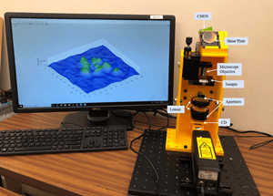 Researchers designed a powerful microscopy system that is inexpensive and easy to replicate because it is made of 3D printed parts and commonly found optical components. The image on the screen shows 3D reconstructed results for a green algae sample. (Image credit: Bahram Javidi, University of Connecticut)
“This new microscope doesn’t require any special staining or labels and could help increase access to low-cost medical diagnostic testing,” said research team leader Bahram Javidi from the University of Connecticut. “This would be especially beneficial in developing parts of the world where there is limited access to health care and few high-tech diagnostic facilities.”
The scientists illustrate their new microscope, which is founded on digital holographic microscopy, in The Optical Society (OSA) journal Optics Letters. The portable instrument creates 3D images with double the resolution of traditional digital holographic microscopy, which is usually carried out on an optical table in a laboratory. Besides biomedical applications, it could also be beneficial for manufacturing, defense, research, and education.
The entire system consists of 3D printed parts and commonly found optical components, making it inexpensive and easy to replicate. Alternative laser sources and image sensors would further reduce the cost, and we estimate a single unit could be reproduced for several hundred dollars. Mass production of the unit would also substantially reduce the cost.
Bahram Javidi, Study Team Leader, University of Connecticut.
From the lab to field ready
A digital camera in traditional digital holographic microscopy records a hologram made from interference between a reference light wave and light emitted from the sample. Next, a computer transforms this hologram into a 3D image of the sample. Although this microscopy method is beneficial for exploring cells without any dyes or labels, it usually necessitates a multifaceted optical arrangement and stable atmosphere free of vibrations and temperature variations that can add noise in the measurements. Therefore, digital holographic microscopes are mostly only seen in laboratories.
The scientists could increase the resolution of digital holographic microscopy past what is possible with even illumination by integrating it with a super-resolution method called structured illumination microscopy. They realized this by producing a structured light pattern with the help of a clear compact disc.
“3D printing the microscope allowed us to precisely and permanently align the optical components necessary to provide the resolution improvement while also making the system very compact,” said Javidi.
Testing the new microscope
The scientists assessed the performance of the system by recording images of a resolution chart and then using an algorithm to rebuild high-resolution images. This revealed that the new microscopy system was able to resolve features as small as 0.775 µm, twice the resolution of traditional systems. Using a light source possessing shorter wavelengths would enhance the resolution a lot more.
More experiments revealed that the system was sufficiently steady to examine variations in biological cells over time, which have to be computed on the scale of a few tens of nanometers. The scientists then established the device’s applicability for biological imaging by obtaining a high-resolution image of green algae.
“Our design provides a highly-stable system with high-resolution,” said Javidi. “This is very important for examining subcellular structures and dynamics, which can have remarkably small details and fluctuations.”
The scientists say that the existing system is ready for real-world use. They propose to use it for biomedical applications such as disease diagnosis and cell identification and will continue their partnership with their global associates to examine disease identification in remote areas with inadequate access to healthcare. They are also involved in further improving the system’s resolution and signal-to-noise ratio without raising the device’s cost.