Nikon Instruments Inc. today introduced a new High Content microscope system featuring speed and flexibility for imaging and managing biological assays utilizing a system based on the classic Nikon Ti inverted microscope and NIS-Elements Software.

This new High Content Microscope system pairs the Ti inverted microscope with NIS-Elements HC Software to provide a dedicated interface for high content acquisition and analysis routines, built on an integrated database for seamless data management and processing.
This system incorporates all of the functionality of the Ti+ Perfect Focus microscope with interchangeable options for high-speed piezo-based autofocusing, plate handling, solid state light source and fast wavelength switching as well as the full range of NIS-Elements camera model support. In addition, magnification and fluorescence filters are exchangeable, extending the imaging possibilities and experimental assay design options.
“Nikon is excited to offer a new avenue for researchers to add high content applications to their imaging without giving up the power and flexibility of their Ti ‘Perfect Focus’ workhorse,” said Stephen Ross, Ph.D., General Manager of Product and Marketing at Nikon Instruments Inc. “There’s a lot to be said for the confidence and ease of a familiar instrument when you’re scaling up the imaging capability within the same system.”
Hallmarks of the new High Content system and NIS-Elements HC package include:
- Streamlined high-speed automated well plate acquisition, data review analysis and management of multiple well plate runs
- Easy step-through plate configuration navigation
- Built-in library of commonly used well plate formats
- Easy routine for labelling wells and their treatments
- Automated alerts through text and email for system updates
- Automatic scheduling for managing the experimental pipeline
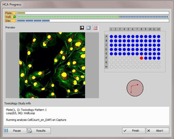
- Real-time viewing of data acquisition and analysis progress for instant inspection and evaluation.
- Displays high content data in a variety of viewing options
- High Content data displayed in a variety of formats to allow for full access to metadata, customizable heat maps, measurement results, generated binary masks, assay results and sample labeling
- Data is easy to filter and export
Nikon’s High Content microscopy system was introduced in the United States at the American Society for Cell Biology Annual Meeting December 15-19, 2012 and available in December 2012.