EMBL Heidelberg researchers used a new microscope for capturing a 3D film of a fruit fly embryo. It was two-and-a-half hours old when it emerged out of the microscope as a larva.
A new microscope enabled scientists to film a developing fruit fly embryo, in 3D, for over 20 hours. ©EMBL/U.Krzic
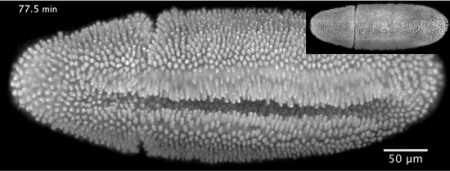
The video was created based on a new microscope developed at Hufnagel’s lab. It was published online in Nature Methods. The video shows the formation of the ventral furrow resulting from the movement of cells on the embryo’s belly, while the other cells move in a convergent extension, starting around the embryo’s rear end through its back (inset). Following an opening within the embryo’s inset, dorsal closure occurs wherein the surrounding cells close the gap.
Using this new microscope named Multi-View SPIM( MuVi-SPIM), researchers can effectively image expedite biological processes in clear, thick samples. The EMBL-developed Selective-Plane Illumination Microscopy (SPIM) technology forms the basis of this microscope. Similar to SPIM, the new microscope sheds a thin film of light on the embryo, where one layer of a sample will be illuminated at a time, generating the image of whole sample involving very low light-induced damage. MuVi-SPIM captures four full images from different angles, without changing the direction of sample. This novel device enables the scientists to combine all the four images into a single 3-D image of high quality.
Hufnagel and his associates were able to rapidly capture a high-quality 3-D image of a fruit fly embryo. Requiring only a few seconds to complete the imaging process, this microscope enables the scientists to resolve the property of each cell or the structures within the cells. This rapid imaging enables Hufnagel and colleagues to record the movements of each nucleus within the embryo all through the first 3 hours of the lifecycle of the embryo, demonstrating the rapid division of nuclei.
The scientists will deploy the new microscope to study the formation of organs and tissues in other model organisms in addition to fruit fly.
Disclaimer: The views expressed here are those of the author expressed in their private capacity and do not necessarily represent the views of AZoM.com Limited T/A AZoNetwork the owner and operator of this website. This disclaimer forms part of the Terms and conditions of use of this website.