Confocal microscopes use a distinct arrangement of apertures to generate an image at the detector that is a small optical slice or segment of the specimen. A sample that is several millimeters thick can be narrowed to a focal field of less than one micron in the z-axis, for example.
The confocal's field of illumination is limited by an aperture, commonly known as a pinhole. The field of view is also restricted by a pinhole conjugate to the lighting point and the first aperture.
This 'confocal' design reduces the amount of out-of-focus light reaching the detector, thereby increasing the signal-to-noise ratio and significantly reducing the image volume.
Modern scanning confocal microscopes employ a laser or a combination of lasers as their illumination source. The scan is performed using carefully regulated galvanometer mirrors that move the diffraction-limited patch of illumination across the field in a raster motion, comparable to a CRT television set operated by a computer.
The dispersed, reflected fluorescence light from the sample is routed to a photomultiplier tube (PMT), and the image is displayed on a computer screen, one point (or pixel) at a time.
One of the most essential characteristics of confocal microscopy is the capacity to optically segment specimens. Confocal optical sectioning enables users to image thick tissue without microtomy and to view living cells, tissues, and organisms with high resolution.
This optical sectioning capability also allows the individual slices/images to be stored on the computer and subsequently reconstructed to display the sample's three-dimensional portrait.
This technology imposes exceptional restrictions on the optical elements employed in fluorescence imaging. A common misconception is that the laser light output of a particular laser produces only one wavelength. In actuality, practically all lasers emit harmonics and/or scatter other wavelengths.
While these secondary lines may be weaker in strength than the primary line, they can still significantly reduce the signal-to-noise ratio. If the fluorescence emission occurs at the same wavelength as the harmonic (or other laser noise), the fluorescent signal can be entirely obscured, rendering it invisible.
Filter Requirements for Confocal Microscopy
Excitation Filters
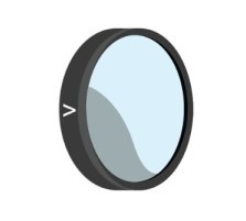
Image Credit: Chroma Technology Corp.
The first optical device in the excitation path is usually a laser clean-up filter, which guarantees that only the desired laser line enters the specimen.
These filters must transmit light efficiently at the laser wavelength while blocking undesired spectral components over a wide range. Clean-up filters are typically low-bandpass filters (∼10 nm FWHM), but diode lasers may require slightly wider designs (20–25 nm FWHM) due to wavelength drift.
Because laser light is collimated and coherent, clean-up filters must meet stringent optical quality standards. These include reduced wavefront distortion and a small wedge to prevent beam deviation and misalignment.
Dichroic Mirrors

Image Credit: Chroma Technology Corp.
Dichroic beamsplitters in confocal systems are chosen for their excellent flatness to retain beam quality and reduce wavefront distortion. In some circumstances, thicker substrates are used to meet transmitted wavefront specifications. These optics must also tolerate intense laser power without degrading.
Emission Filters
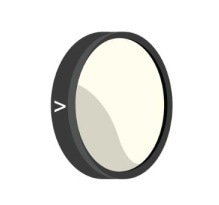
Image Credit: Chroma Technology Corp.
The primary function of emission filters in confocal systems is to block excitation light while passing fluorescence.
Because laser lines can be exceedingly bright, the blocking needs may reach OD8 for certain wavelengths. Although the spectral range of blocking is often narrower than in widefield fluorescence, the required attenuation at the laser wavelength is much greater.
Note About Filter Sets for Confocal Microscopy
Filter sets for confocal systems are often constructed around available laser lines rather than fluorescence peak excitation. As a result, fluorophore choice is frequently limited by the available laser wavelengths. Polychroic beamsplitters can be used to handle several laser lines in multicolor imaging systems.

This information has been sourced, reviewed, and adapted from materials provided by Chroma Technology Corp.
For more information on this source, please visit Chroma Technology Corp.