Total internal reflection fluorescence (TIRF) microscopy reduces background fluorescence while increasing the signal-to-noise (S/N) ratio in the produced images. This is accomplished by using light's capacity to generate an evanescent wave (or field) within a very narrow spatial range beyond the interface of two substrates with different refractive indices.
Principle of Evanescent Wave Excitation
In practice, this involves imaging a specimen in close contact with a glass slide or tissue chamber. When the angle of incidence exceeds the critical angle, the refractive-index imbalance produces an evanescent field with frequency components that match those of the incident light.
In simple terms, a fluorescent molecule that would ordinarily absorb light at 488 nm can be 'activated' by the electromagnetic field produced by a 488 nm laser (or other monochromatic source) and reflected off of the lower refractive index material.
Because the intensity of this evanescent field decreases exponentially with distance from the contact, the fluorescence signal will appear within less than 100 nanometers of the surface/sample interface.
This efficiently restricts excitation to the interface, lowering the z-axis signal while greatly boosting the fluorescence signal-to-noise ratio. This implies that the molecules of interest must be within the same restricted range as the evanescent field.
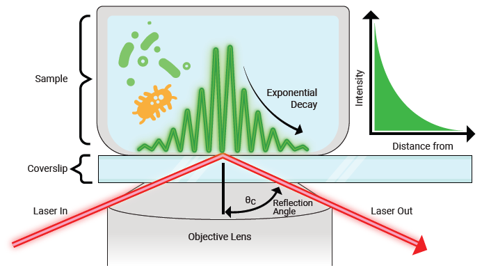
Figure 1. Evanescent field generated by total internal reflection, showing exponential decay in intensity. Image Credit: Chroma Technology Corp.
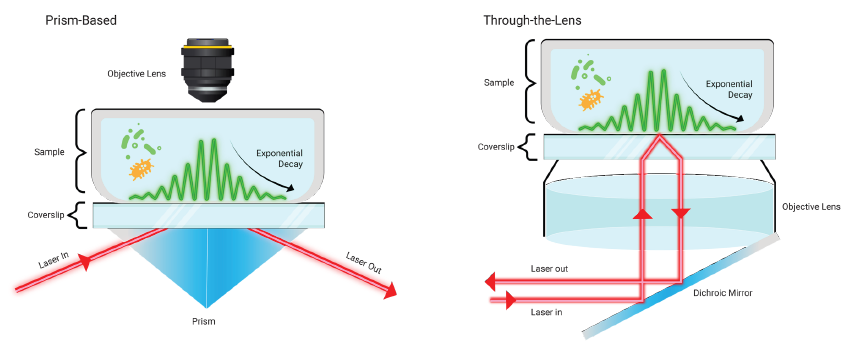
Figure 2. Two approaches to TIRF microscopy: prism-based excitation (left), which uses external beam delivery, and through-the-lens excitation (right), which directs the laser through the objective for improved usability and system integration. Image Credit: Chroma Technology Corp.
From Prism-Based TIRF to Through-the-Lens Systems
TIRF has been used for decades in a variety of applications. The earliest TIRF microscopy experiments used prisms and beam-steering optics to transmit the excitation laser or light to the sample via an inverted microscope configuration.
Researchers can now use 'through-the-lens' microscope systems with high-numerical-aperture objective lenses to reduce alignment time and enhance system safety by using an isolated beam path.
Increased Optical Demands in Through-the-Lens TIRF
The 'through-the-lens' approach to TIRF creates great demands on optical filters since the reflected excitation beam re-enters the objective and travels backward through the emission channel. As a result, the emission (barrier) filter has to tolerate exceptionally high excitation energy, as well as high-angle scatter and reflections.
In standard laser-based fluorescence microscopy, the emission filter must block excitation wavelengths up to OD 6 (OD = -log[%T]). However, in TIRF, blocking must surpass OD 8. Many typical spectrometers cannot detect this amount of deep blockage.
To meet these specifications, Chroma employs proprietary coating designs, blocking mechanisms, and high-quality optical substrates to provide OD 8 performance even at the high angles inherent in TIRF illumination.
Reflected Wavefront Distortion (RWD) Requirements
Aside from blocking performance, the dichroic mirror in a TIRF system must have extremely low reflected wavefront distortion (RWD).
TIRF systems need less than 1/2 wave of distortion to maintain the integrity and uniformity of the evanescent field, unlike ordinary laser-scanning confocal microscopes that require fewer than two waves per inch.
Achieving this level of flatness is difficult because the coating deposition process generates mechanical forces that can deform even high-quality substrates.
Extremely thin or oversized substrates are particularly vulnerable. This makes substrate thickness, coating process, and stress-compensation procedures important for consistent TIRF performance.

This information has been sourced, reviewed, and adapted from materials provided by Chroma Technology Corp.
For more information on this source, please visit Chroma Technology Corp.