Electron Microscopy for tool mark analysis has revolutionized forensic investigations, offering unparalleled precision and clarity in examining minute surface details. This article will explore the application and significance of electron microscopy in tool mark analysis, revealing its potential for extracting crucial insights into the development and characteristics of tool marks.
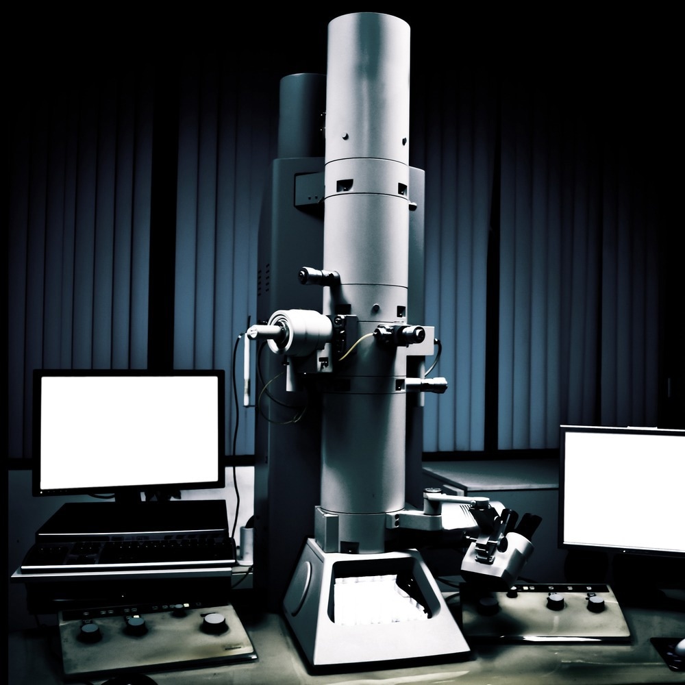
Image Credit: Pan Xunbin/Shutterstock.com
Overview of Toolmark Analysis and Its Importance in Forensic Investigations
Toolmark analysis is a vital forensic technique to identify tools during forensic investigations. It involves studying the unique marks left by various tools and comparing them to identify potential matches. These bookmarks provide valuable information about the characteristics of the tools, including shared features and unique attributes.
Forensic experts analyze the damage impressions and manufacturing marks to establish a tool's distinctiveness and link it to a crime. This analysis helps investigators gather essential evidence and accurately identify the tools involved.
The Significance of Electron Microscopy for Tool Mark Analysis
The introduction of the electron microscope into forensic investigations occurred soon after its commercial debut in the 1940s. Since then, it has played a prominent role in the identification, topographic comparison, and interpretation of physical evidence, addressing aspects that cannot adequately handle by light microscopy.
In the electron microscope, image formation primarily relies on the emission of secondary electrons from the specimen surface, providing a high resolution (>10 nm) and a remarkable depth of field (300-500 times that of a light microscope) across a wide range of magnifications.
Forensic investigators have recognized and harnessed these advantages, particularly in tool mark examination, allowing them to visualize specimens as small as 0.02 pm, surpassing the capabilities of optical microscopy. This closer inspection has proven invaluable in identifying and distinguishing morphology, texture, and elemental composition characteristics.
Another significant advantage of electron microscopy is its non-destructive nature, ensuring the preservation of the tool mark during analysis. This preservation is crucial for further examination or comparison, contributing to thorough forensic investigations.
How is Toolmark Analysis Conducted Using Electron Microscopy?
The tool mark analysis begins with capturing high-resolution digital images of marking patterns using an electron microscope. These images are then analyzed to extract relevant features, such as characteristic damage impressions and manufacturing marks.
By optimizing the imaging conditions, electron microscopy reveals fine topographic details crucial for tool mark identification. The acquired images are further processed using an optoelectronic correlator device, which performs numerical and optical analyses of the feature data.
This comprehensive approach transforms the two-dimensional striation patterns into one-dimensional barcode information, preserving the original data and enabling robust comparisons. The correlation measures obtained through this method serve as objective criteria for identifying the tools involved.
Which Patterns Are Examined Using Electron Microscopy for Tool Mark Analysis?
Electron microscopy for tool mark analysis enables forensic experts to identify and compare possible identification marks, scrape marks, and cut surfaces exhibiting fine striations and irregularities.
Indentation Marks
Indentation marks occur when a tool is pressed against a surface without sliding, resulting in surface irregularities that outline the tool's surface. Examples include firing pin impressions, often encountered when identifying cartridges in firearms cases.
While these marks are visible under an optical microscope, comparing them to trial impressions can be challenging, especially when the cartridge is punched. The electron microscope has proven valuable in facilitating such comparisons.
Scrape Marks
Scrape marks, characterized by lines or striations, result from tools such as chisels, knives, axes, pliers, scissors, or screwdrivers sliding across a surface. These marks are distinct and can be compared to trial scrape marks using an optical microscope.
However, striations produced on cut surfaces are more complex than those created by surface scratching, making them challenging to compare under an optical microscope.
Cut Surfaces
Cut surfaces, resulting from the action of a single wedged tool such as knives, scissors, or chisel, exhibit a complex surface structure of fine striations and irregularities.
Although the two surfaces may have different inclinations and smoothness, they will exhibit the same striations due to the common edge of the tool. By regenerating and matching these striations under an electron microscope, it is possible to confirm that the two parts were cut from the same material.
Future Outlooks and Advancements in Electron Microscopy for Tool Mark Analysis
Electron Microscopy for tool mark analysis has revolutionized forensic investigations by enabling high-resolution imaging and feature extraction from tool marks. It has proven particularly effective in differentiating characteristics like morphology, texture, and elemental composition in firearm markings.
New developments in electron microscopy will also allow for dynamic analysis of tool marks, tracking tool movement during mark creation, and aiding crime scene reconstruction and event determination. In addition, aberration-corrected electron microscopy and other advanced imaging techniques will offer even higher-resolution images, refining forensic comparisons and potentially introducing novel tool mark identification methods.
Electron microscopy's application is expanding beyond traditional materials such as metal, plastic, wood, and glass, as efforts are underway to analyze tool marks on textiles and paper. This broadens the scope of tool mark analysis, potentially leading to new methods for criminal identification.
More from AZoM: How is Spectroscopy Used to Analyze Fire Debris?
References and Further Reading
Basu, S., & Millette, J. R. (2012). Electron Microscopy in Forensic, Occupational, and Environmental Health Sciences. Springer Science & Business Media. https://books.google.com.pk/books/about/Electron_Microscopy_in_Forensic_Occupati.html?id=5k0yBwAAQBAJ&source=kp_book_description&redir_esc=y
Demoli, N., Šariri, K., Stanić, Z., Maštruko, V., & Milat, O. (2004). Toolmarks identification using SEM images in an optoelectronic correlator device. Optik, 115(11-12), 487-492. https://doi.org/10.1078/0030-4026-00404
Kumar, S., Saxena, G., & Gautam, A. (2021). Forensic Analysis and Interpretation of Tool Marks. IntechOpen. https://www.doi.org/10.5772/intechopen.98251
Nanoscience Instruments. (2023). What Role Does SEM Play in Trace Evidence Analysis? [Online]. Available from: https://www.nanoscience.com/blogs/what-role-does-sem-play-in-trace-evidence-analysis
Disclaimer: The views expressed here are those of the author expressed in their private capacity and do not necessarily represent the views of AZoM.com Limited T/A AZoNetwork the owner and operator of this website. This disclaimer forms part of the Terms and conditions of use of this website.