Plants, it is fair to say, may be aspects of our world that many of us take for granted. As far as we know, plants have in some form always existed – and so we expect that they always will. But is that the case?

Image Credit: Renishaw plc – Spectroscopy
Like all species on our planet, plants evolve, adapting to increasingly urban areas, changing habitats and climates, and environmental shifts. The current rapid pace of human-driven changes can pose a threat to some plants - which in the past have been able to gradually adjust to external factors. As plants provide 80% of our food and 98% of the oxygen we breathe, they are vital to us, significantly impacting our economy and environment [1]. 2020 was therefore declared by the United Nations as the International Year of Plant Health (IYPH). [2]
There is a range of factors that actively influence plant health, growth, and resistance to disease. It is vital that plants are monitored and that any changes taking place are detected - ideally before the visible signs of them occur. Suitable analytical techniques are therefore needed for this.
Raman spectroscopy is an ideal technique for this, as it is non-destructive, offers highly specific chemical information, and can analyze samples in water. This allows researchers to measure biological processes both ex vivo and in vivo.
In this article, research on plants is demonstrated, which is performed using Renishaw's inViaTM confocal Raman microscope. This research grade instrument is extremely well-suited to the analysis of plant tissue. It can image botanical samples with a high spatial resolution and chemical specificity, thanks to its advanced hardware and software.
From Kernel to Wood
Grains
One of the main food sources of our diet, cereal grains are integral to modern and ancient societies. Their industrial processability is directly influenced by their structure and chemical composition, and this also influences how they are broken down in both animal and human digestion systems.
The spatial distribution of components in a cross-section of a kernel can be revealed by Raman imaging. Figure 1 depicts the Raman image of a wheat grain cross-section. Specific sections of the kernel structure can be easily distinguished, including the aleurone layer cell (center), starchy endosperm (left bottom corner), aleurone-pericarp border (right top corner). [3] We can understand the effect of external factors on plants and their seeds by interpreting distributions such as these. This supports the development of more robust grain varieties.
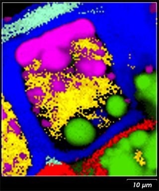
Figure 1. Raman chemical image of a wheat grain cross section, showing arabinoxylan (blue), β-gucan (red), protein (yellow), lipids (magenta), starch (green) and xylan (cyan). Image Credit: Renishaw plc – Spectroscopy
Metabolites
If we can track the effect of environmental, nutritional and mechanical stresses on a plant's tissues we can reveal their impact on the plant's development and health. [4]
Within a plant's tissues and organs (roots, wood, stems, bark, leaves, seeds, etc.), different types of metabolites can form. We can gain insights into how plants adapt to environmental changes, and how their growth will be affected by climate change if we can monitor and understand metabolite production.
One key metabolite is calcium oxalate. Calcium oxalate protects against herbivores, regulates calcium levels in tissues, strengthens tissues, detoxifies heavy metals, and helps in the gathering of light for photosynthesis. A combination of genetic and environmental factors influence and define its amount, crystal shape, size and function. [5]
Raman spectroscopy can easily assist in the study of spatial distribution of calcium oxalate crystals in plant tissues and organs. An optical image of an English oak leaf, and the corresponding Raman image are shown by Figure 2. The lignin content, with its highest content at the compound middle lamella (CML) and cell corners (CC) is shown by the Raman image. The large calcium oxalate metabolite crystals are present in the corners of the leaf cells (Fig. 2B; cyan). Raman spectroscopy can also be used because of its high chemical specificity to study the hydration state of calcium oxalate.
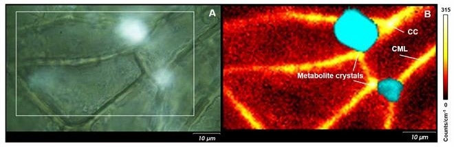
Figure 2. (A) Optical image of a cross section of an English oak leaf; (B) corresponding Raman image showing lignin (yellow/orange), and calcium oxalate (cyan). Image Credit: Renishaw plc – Spectroscopy
Wood
Trees are understandably vital to our way of life. In addition to producing oxygen for us to breathe, they give space for wildlife to live and reproduce. Trees are also crucial to our economy, having been used for centuries in the production of furniture, paper, and buildings.
Our ability to predict the mechanical durability and resistance to biological attack and decomposition hinges on our understanding of the complex structure of wood tissues.
Wood is primarily made up of hemicelluloses, cellulose, and lignin. The high specificity of Raman spectroscopy can be used to study the organization and structure of wood cell walls. The presence of the secondary metabolites called extractives influences their resistance to decomposition and biological attack.
The distribution of lignin in a cross-section of Scots Pine is shown by Image (A) of Figure 3. Lignin is concentrated at the compound middle lamella (CML; middle lamella + adjacent primary walls) and cell corners (CC), with a higher concentration than is shown in the sublayer of the secondary cell wall (S2). A polarized laser was used to reveal the variation in cellulose orientations generated Image B. Two small layers next to the CML, with a high intensity of the cellulose band show a sublayer of the secondary cell wall (S1).
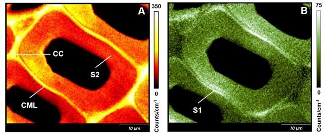
Figure 3. Raman images of a cross section of Scots pine wood. (A) shows the distribution of lignin, (B) the orientation of cellulose. Image Credit: Renishaw plc – Spectroscopy
Bright Future
Each year, the pace of the observed changes in the environment quickens. This is also true for plants. Faster and simpler analytical methodologies are required by this successful approach to the new reality. Raman imaging, in this context, plays an increasingly important role in the study of plant organs and tissues. This is because it is enormously versatile, providing chemical and spatial analysis, images, and information on molecular orientation.
Raman spectroscopy can offer a key element in this new approach to the studies on plant health. This can provide missing piece of the puzzle, in which a crucial role will be played by the inVia confocal Raman microscope.
Acknowledgments
Produced from materials originally authored by Anna Lewandowska, Application Scientist from Renishaw.
References
- https://www.yearofplanthealth.co.uk/why-plants-are-important
- http://www.fao.org/plant-health-2020/home/en/
- A.-S. Jskelinen, U. Holopainen-Mantila, T. Tamminen, T. Vuorinen, J. Cereal Sci. 57 (2013) 543-550
- H.J. Butler, M.R. McAinsh, S. Adams, F.L. Martin, Anal. Methods, 7 (2015) 4059-4070
- M.N. Islam, M. Kawasaki, Plant Prod. Sci. 17 (2014) 13-19

This information has been sourced, reviewed and adapted from materials provided by Renishaw plc - Spectroscopy.
For more information on this source, please visit Renishaw plc - Spectroscopy.