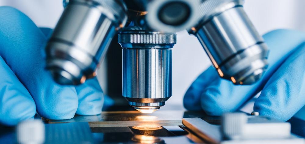
Cells are very delicate and easily damaged, particularly when used as a bioink in additive manufacturing to create living structures such as bioimplants. A new microscopic imaging approach allows scientists to see how this harm might occur and could lead to better bioimplants, as well as improved disease modeling and drug screening.
A study developed by the University of Birmingham sheds light on how cells become damaged, making way for improved bioimplants. Image Credit: Konstantin Kolosov/Shutterstock.com
Additive manufacturing has transformed research and manufacturing worldwide; it automatically creates a predefined object layer-by-layer using computer-aided design and a special ink – in the case of bioimplants, these bioinks contain fragile cells, biomolecules, and materials.
Additive Manufacturing for Cell Damage
In the medical field, additive manufacturing has been used to create bioimplants such as porous bone implants, prosthetics, wearable biosensors, and drug delivery systems. These implants aim to fix, support, reproduce or improve the function of human tissues by integrating them into the body.
Demand for patient-specific devices has grown in recent years, thanks in part to an aging population, and a shortage of donor organs for medical treatments. Such bio-printed structures can also be matured in vitro to create models of tissues and organs for disease modeling, drug screening, or regenerative medicine.
Extrusion-based bioprinting is widely used in biofabrication because it is quick, precise and simple, and delivers a living object by extruding a hydrogel – which acts as a support matrix - via a thin capillary tube with a diameter between 50 µm to 1 mm onto the desired template architecture.
Novel Imaging Technique
Despite the benefits of the technique, cells squeezing through narrow tubes during the bioprinting process can be easily damaged. Their cell viability, phenotype (observable traits), and functionality can all be affected. The damage can be compounded by the narrower capillaries, large flow rates, and the high viscosity hydrogels needed for quick printing with ample resolution and shape integrity.
A new microscopic imaging approach developed by scientists from the University of Birmingham sheds light on how this damage occurs and could lead to improvements in the 3D printing process.
“We used light sheet fluorescence microscopy (LSFM) to mimic the portion of the extrusion bioprinting process in which cells are most likely to be damaged,” says Dr. Gowsihan Poologasundarampillai, Fellow in Biomaterials and Bioimaging at the School of Dentistry.
The new method shines a blade of light in the bioink as it flows inside the narrow printing tube, and illuminates a range of hydrogel-cell behavior and damage patterns, which depend on the extrusion speed and properties of the tube utilized during printing.
“Printing parameters and hydrogel flow behavior can determine the degree to which cells are mechanically damaged as they pass through the capillary,” explains Poologasundarampillai, lead author of the study published in Bioprinting.
“Direct observation of the hydrogels and suspended cells during the printing process will help shed light on the conditions leading to cell necrosis,” he adds.
The novel technique could provide an improved understanding of bioink flow dynamics and how cells move, enabling better complex capillary designs and processes.
“We’ve demonstrated the power of LSFM-based imaging to give new insights on cell and fluid movements and flow patterns during extrusion using different bioinks,” says Poologasundarampillai.
This information on cell dynamics was lacking from previous research on flow behavior and mechanical stress on cells during printing, he adds, often leading to faulty assumptions about fluid behavior and flow modeling.
Going Forward
Poologasundarampillai believes the novel imaging approach could be further exploited to improve the outcomes of 3D bioprinting, and the researchers applied their new imaging technique to study the real-time flow of bioink formulations that showed constant and shear thinning viscosities through a capillary.
This new real-time imaging technique offers new insights on cell and fluid movements and flow patterns during extrusion, and could result in the development of fresh capillary designs featuring complex structures and surface chemistry to modulate cell viability during extrusion bioprinting.
Optimal bioink design maximizes cell survival and the structural integrity of the resulting object. This method could also aid the design of novel bioinks with better flow properties, enhancing cell viability and the overall success of bioprinting.
References and Further Reading
Poologasundarampillai, G. et al. (2021) Real-time imaging and analysis of cell-hydrogel interplay within an extrusion-bioprinting capillary: Bioprinting - https://doi.org/10.1016/j.bprint.2021.e00144. Accessed 8 June 2021.
University of Birmingham (2021) New imaging technique could lead to better bio-implants for patients: University of Birmingham - https://www.birmingham.ac.uk/news/latest/2021/05/better-bio-implants-for-patients.aspx. Accessed 8 June 2021.
Liu, G. et al (2020) Development of Bioimplants with 2D, 3D, and 4D Additive Manufacturing Materials: Engineering - https://doi.org/10.1016/j.eng.2020.04.015. Accessed 8 June 2021.
Disclaimer: The views expressed here are those of the author expressed in their private capacity and do not necessarily represent the views of AZoM.com Limited T/A AZoNetwork the owner and operator of this website. This disclaimer forms part of the Terms and conditions of use of this website.