Image Credit: Motortion Films/Shutterstock.com
Microscopy techniques are used in many fields. High-powered and high-resolution microscopy methods – such as the electron microscope – can now be used to see many things in more detail. When it comes to the food industry and analyzing food samples, microscopy and imaging methods are ideal because they enable the person to physically see the food sample, rather than having to analyze it via a numerical output (although many images nowadays can be further converted in mathematical and statistical data to provide a deeper understanding of what’s happening in the image/sample).
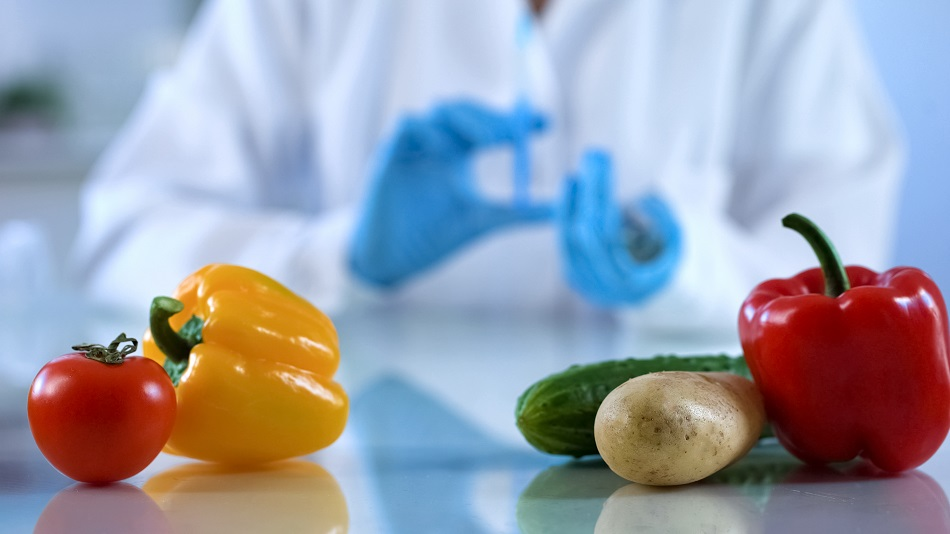
Microscopes are used throughout the food industry and their use has been growing as scientists now analyze a wide variety of foodstuffs and contaminants within foodstuffs – from particles of food to food fibers, to food pathogens and many more in between. These analyses are important as they are not only used to show if the foodstuff has the right rheological texture and structure, they are also used to see the microbiological mechanisms occurring within the foodstuff, which can be used as an indicator for how long it will be before food will spoil.
Microscopes and imaging methods can be used with a wide variety of samples, from pure liquids and solids to more complex mixtures such as emulsions. The techniques available to researchers range from the simple benchtop optical microscope to high-powered electron microscopes, and even magnetic imaging resonance (MRI) microscopes.
Electron Microscopy (EM)
Of all the electron microscopy (EM) techniques, scanning electron microscopy (SEM) is the most widely used and provides the highest image resolution of the methods which are used in a commercial and industry-focused setting. SEM is widely used to image the surface of the sample, but to analyze the internal structure, transmission electron microscopy (TEM) is used in conjunction with SEM. SEM and TEM use a beam of high energy electrons to interact with the sample where the detection of these electrons is used to generate an image of the sample surface. For SEM, the electrons interact and scatter with the sample, whereas they pass through the sample in TEM.
With an EM, it is possible to go down to 1 nm resolution (much greater than optical microscopes) so it has become a way of seeing foodstuffs in much more detail, as well as seeing some of the finer structures which contribute to the texture and sensory feel of the foodstuff. Despite their many advantages, the one disadvantage of EM techniques is that they can’t be used with liquid samples due to the analysis being performed in a vacuum. This means that the samples need to be frozen or dried, however, this also means that that how the different drying and freezing processes affect foodstuffs can be easily studied. Aside from drying processes, EM methods can be used to investigate the structure and properties of many different foodstuffs, from oils to fruits, meats, vegetables, and many more in between.
Optical Microscopy
Optical microscopes are the conventional benchtop microscopes that many laboratories have and use photons of light to illuminate the sample – where the user can see the sample either through the eyepiece or via an attached camera. Optical microscopes attached to video cameras can also capture live videos and is particularly useful for capturing how fluidic foodstuffs behave (as fluidic foodstuffs can be imaged unlike in EM). Fluorescent markers can also be added to foodstuffs to highlight certain areas of interest – much like in many medical fields – and to see if there are any contaminants in the sample. Overall, optical microscopy is widely used as you can see the foodstuff in a bit of detail, it can be used with a wide variety of samples and it is a cheap and quick way of performing an initial analysis. However, it lacks the resolution and power that EM microscopes have, but that is not always required for some analyses.
Confocal microscopy is a variation on optical microscopy that has a slightly better resolution than many conventional optical microscopes. This is brought about by a spatial pinhole being placed in the focal plane of the lens. This small adjustment enables the confocal microscope to see the structure of foodstuffs in a higher resolution, to see different types of fluorescence, and enables the food’s properties and structures to be reconstructed into a 3D image. It is a more advanced technique than most optical microscopes and has become useful for analyzing the molecules involved with flavor, but its resolution is still not as high as electron microscopes. However, like many of the microscopy techniques, it has its distinct benefits and uses.
Magnetic Resonance Imaging (MRI)
MRI is not a widely used imaging technique due to its size, cost and need for specialized operators. The analysis of foodstuffs tends to err on the cheaper side of analytical instruments than the medical industry does due to budgets and consumer demand for lower-priced products. However, from a scientific point of view, MRI can be used to look at foodstuffs because it can provide a high resolution and is non-invasive. MRI methods that have been applied to foodstuffs have looked at cereals, vegetables, meats, and dairy products with a resolution of around 100 microns. MRI could be used in processing environments to study fruit ripening, drying processes, thermal processing, food growth and food maturing process in real-time, but whether its commercial use will still be limited by its cost in the future remains to be seen.
Sources and Further Reading
Disclaimer: The views expressed here are those of the author expressed in their private capacity and do not necessarily represent the views of AZoM.com Limited T/A AZoNetwork the owner and operator of this website. This disclaimer forms part of the Terms and conditions of use of this website.