The alpha300 Ri inverted confocal Raman imaging microscope offers 3D chemical characterization from a different angle. Its inverted beam path maintains all the functionality of WITec’s alpha300 R confocal Raman imaging microscopes while providing new possibilities in access and handling.
A clear advantage when working with oversized samples and aqueous solutions is the ability to visualize and analyze samples from below.
Studies in the fields of geosciences, biomedicine, and life sciences will benefit from the flexibility and consistency provided by the design of the alpha300 Ri microscope.
Key Features of the alpha300 Ri
- Non-destructive imaging method: Sample staining or other specialized preparation is not required
- Inverted beam path enables liquid sample holders to be positioned on the fixed plane of the stage for rapid and repeatable measurements
- Large samples that would be difficult to analyze using a conventional microscope objective turret can be accommodated by the alpha300 Ri’s stage
- Environmental enclosures and other accessories can be easily integrated
- Includes all the exclusive and proven imaging and spectroscopy capabilities of the WITec alpha300 R range
- Can be used with other microscopy methods such as fluorescence, phase-contrast, and differential interference contrast (DIC)
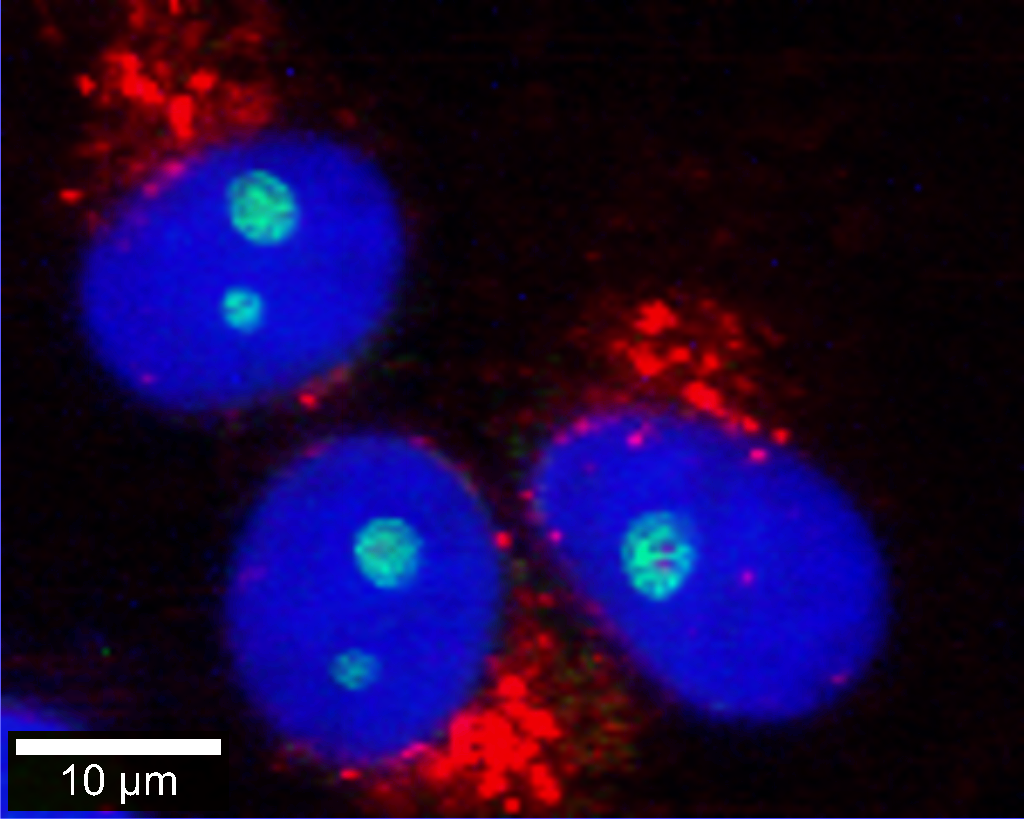
Correlative Raman - fluorescence microscopy image of eukaryotic cells. Nuclei were stained with DAPI (blue). Endoplasmic reticulum (red) and nucleoli (green) were identified by their Raman signals. Image Credit: WITec GmbH
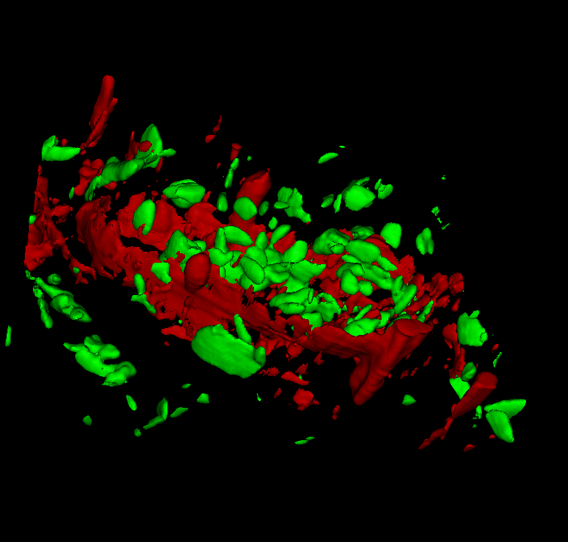
3D Raman measurement of banana pulp. Starch grains (green) and cell wall components (red) are clearly visible. Image Credit: WITec GmbH