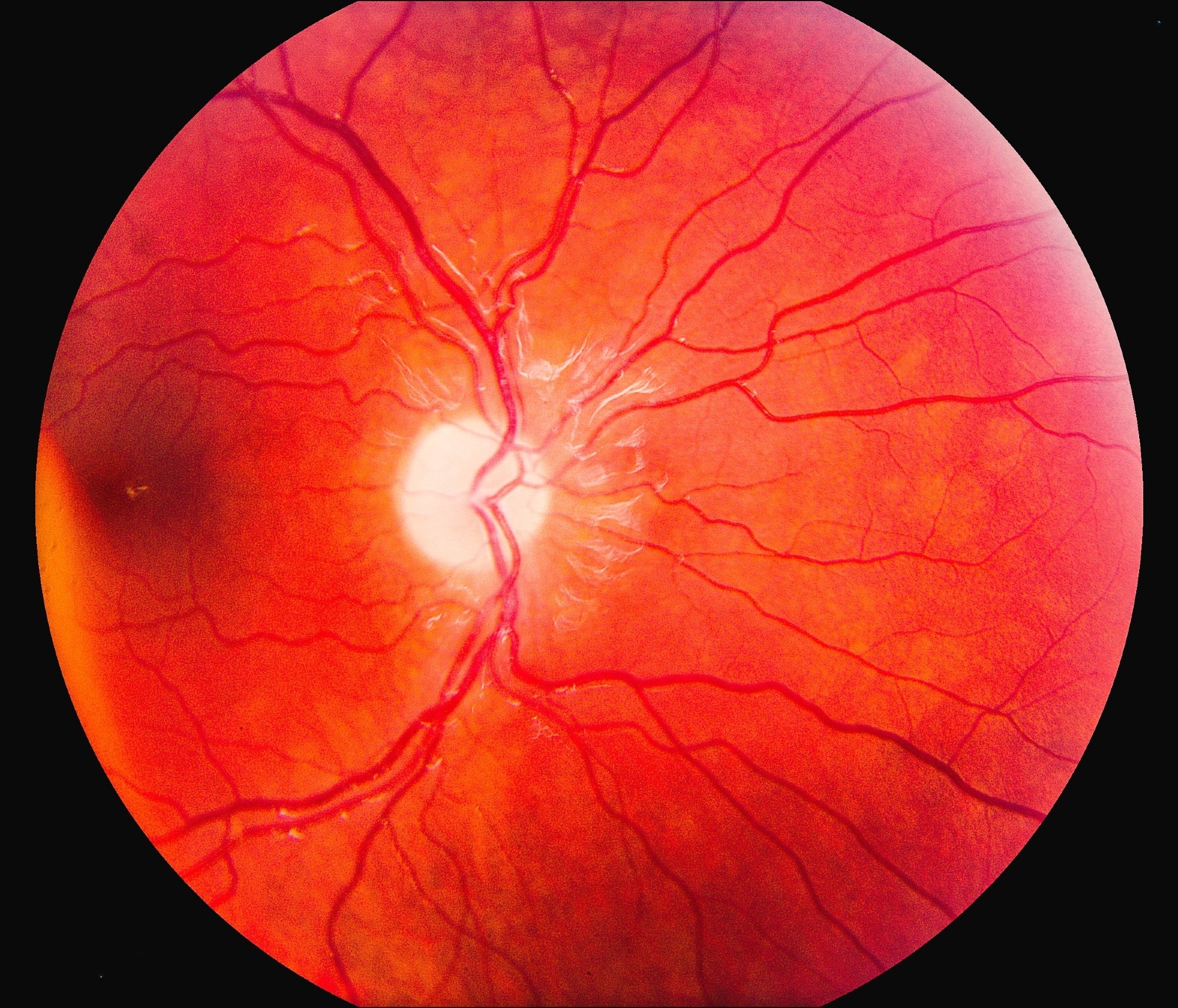
Johns Hopkins University researchers and collaborators have created a digital refocusing technique for retinal imaging that eliminates manual lens adjustments. The method, published in 2024 in npj Digital Medicine and arXiv, uses computational processing after image capture to maintain focus across different retinal layers. Clinical testing with over 1,800 participants showed reduced reliance on technician input and more consistent image quality for diabetic retinopathy and glaucoma assessment.1, 2

Image Credit: James Ebanks/Shutterstock.com
Background
Retinal imaging supports early detection and monitoring of diabetic retinopathy, glaucoma, and age-related macular degeneration. Diabetic retinopathy affects over approximately 103 million people globally, with many facing potential vision loss, making dependable imaging necessary for screening and patient monitoring.3
Current methods like fundus photography and optical coherence tomography (OCT) provide diagnostic value but face accessibility and image quality constraints. Operators frequently need to manually adjust focus for individual eye anatomy, refractive errors, or accommodation differences. This operator dependence creates variability and increases imaging errors.4
These problems are particularly evident in low-resource environments where trained staff and equipment access is limited. Mobile screening programs, used to reach underserved populations, often struggle with image quality consistency. Standard autofocus systems can fail with poor pupil dilation, ocular opacities, or patient movement. Research indicates 10–15% of retinal images need to be retaken due to focus problems, extending exam times, increasing costs, and causing patient inconvenience.5
Download the PDF of the article
Key Findings
The primary innovation involves digitally adjusting focus across retinal layers after images are captured, rather than using mechanical lens movements during acquisition.
The Johns Hopkins team developed a diffuser-based system that eliminates manual lens adjustments for accommodation or refractive error compensation. Lead researcher Corey Simmerer reported computational refocusing across a ±12 diopter range, substantially wider than the typical 2–3 diopter range of conventional autofocus systems.1
Parallel research examined self-imaging OCT (SI-OCT), which performs automated three-dimensional macular imaging without operator adjustment. Testing with 1,822 participants found that SI-OCT integration into screening protocols increased diabetic macular edema detection compared to fundus photography alone.2
Additional studies investigated adaptive optics systems using closed-loop focus shifting with pyramid wavefront sensors. These systems correct ocular aberrations during image acquisition and maintained stable focus in diabetic retinopathy patients, supporting consistent image quality.6
Methodology
Digital refocusing has been implemented through two technical approaches. Phase-mask encoding places a holographic diffuser at the ocular–pupil conjugate, creating a specific point-spread function that captures focus information throughout the image. Computational methods then reconstruct focused images at selected depths using this encoded information.1
The second approach is volumetric computational processing. In this case, self-imaging OCT (SI-OCT) is an example, capturing three-dimensional macular data and applying algorithms to digitally align and refocus retinal layers.2
Here is an easy way to think about it: instead of capturing a perfectly focused image at the moment of the shot, the system intentionally takes a slightly blurred photo, but also collects extra data about the blur itself. Later, a computer processes that information to “refocus” the image, as if the lens had been properly adjusted at the time of capture.
Validation studies have confirmed the clinical potential. The SI-OCT trial measured diagnostic accuracy, acquisition time, and image quality across nearly 2,000 participants, showing strong utility in diabetic eye disease screening.2 Similarly, adaptive autofocus algorithms with image-based sharpness metrics have achieved 90% success rates across 80 human cases while minimizing acquisition time.7
Implications and Applications
The most immediate benefit is a reduction in imaging errors and technician training requirements. Currently, technicians may need months of practice before consistently capturing diagnostic-quality images. With digital refocusing, even minimally trained operators can acquire usable results, expanding access in primary care settings and rural regions.
Telemedicine stands to benefit significantly. Many mobile screening programs have struggled with high dropout rates due to poor image quality or repeat examinations. By enabling reliable images from the first capture, digital refocusing reduces these risks and supports efficient remote diagnosis.
Cost-effectiveness modeling suggests additional advantages. A recent analysis of SI-OCT integration showed favorable ratios by lowering training costs and improving diagnostic yield.2 Performance improvements are substantial: diffuser-based systems achieve wide fields of view and computational refocusing ranges exceeding traditional devices by 4–6 fold, with processing times approaching real-time performance.1
This technology could also accelerate the integration of artificial intelligence into ophthalmology. AI models for disease detection require large datasets of consistent, high-quality images. By reducing variability from technician errors, digital refocusing can provide cleaner data streams for training and real-world deployment.11,12
Handheld and smartphone-based fundus cameras may be the next frontier. Incorporating digital refocusing into compact devices could make advanced retinal imaging possible in rural areas and low-income countries, where infrastructure for standard tabletop systems is limited. Early studies suggest handheld cameras can already approach the diagnostic accuracy of larger devices.4, 8
Limitations and Future Research
Despite its promise, digital refocusing has limitations. Processing remains computationally intensive, which may restrict real-time performance in current clinical workflows. Further optimization is needed to balance speed with image quality.
Resolution trade-offs can occur, as increasing the refocusing range may reduce fine detail, though recent advances have mitigated this issue.9 The system also performs best with dilated pupils and may be less effective in patients with significant ocular opacities or poor cooperation.
Patient-specific challenges remain. While ±12 diopters cover most refractive errors, extreme cases may still require conventional focusing methods. More validation is also needed in pediatric populations and patients with unusual ocular anatomy.
Future research is focusing on integrating artificial intelligence for automated disease detection, extending refocusing to ultra-widefield imaging, and combining computational methods with adaptive optics to maximize resolution and range.6, 10 Researchers also anticipate applications beyond ophthalmology, such as intraoperative imaging and surgical guidance.
Conclusion
Digital refocusing is an emerging approach in retinal imaging that reduces the need for manual adjustments and may help broaden access, lower training requirements, and improve consistency in image acquisition. Some challenges remain, particularly in relation to processing demands, resolution trade-offs, and validation across diverse patient groups. Computational methods are likely to play a growing role alongside traditional optical focusing, and further research will clarify how these techniques can be integrated into clinical practice to support screening and treatment.
Ready to bridge human and artifical vision? Read on here
References
- Simmerer, C., Morakis, M. M., Gomez-Perez, L., Liu, T. Y. A., & Durr, N. J. (2024). In vivo fundus imaging and computational refocusing with a diffuser-based fundus camera. arXiv. https://doi.org/10.48550/arxiv.2406.00122
- Liu, Z., Han, X., Gao, L., & et al. (2024). Cost-effectiveness of incorporating self-imaging optical coherence tomography into fundus photography-based diabetic retinopathy screening. npj Digital Medicine. https://doi.org/10.1038/s41746-024-01222-5
- Zhang, S., Webers, C. A. B., & Berendschot, T. T. J. M. (2024). Computational single fundus image restoration techniques: A review. Frontiers in Ophthalmology. https://doi.org/10.3389/fopht.2024.1332197
- Lu, L., Ausayakhun, S., Ausayakuhn, S., & et al. (2023). Diagnostic accuracy of handheld fundus photography: A comparative study of three commercially available cameras. PLOS Digital Health. https://doi.org/10.1371/journal.pdig.0000131
- Diogenes, I., Restrepo, D., Ribeiro, L. Z., & et al. (2025). Impact of mydriasis on image gradability and automated diabetic retinopathy screening with a handheld camera in real-world settings. medRxiv. https://doi.org/10.1101/2025.01.02.25319898
- Brunner, E., Kunze, L., Laidlaw, V., & et al. (2024). Improvements on speed, stability and field of view in adaptive optics OCT for anterior retinal imaging using a pyramid wavefront sensor. Biomedical Optics Express, 15. https://doi.org/10.1364/boe.533451
- Liu, Z., Qiu, S., Cai, H., Wang, Y., & Chen, X. (2023). Enhancing autofocus in non-mydriatic fundus photography: A fast and robust approach with adaptive window and path-optimized search. Applied Sciences, 14(1). https://doi.org/10.3390/app14010286
- Tan, C. H., Kyaw, B. M., Smith, H., & et al. (2020). Use of smartphones to detect diabetic retinopathy: Scoping review and meta-analysis of diagnostic test accuracy studies. Journal of Medical Internet Research. https://doi.org/10.2196/16658
- Thomas, L., Kulkarni, C., & Kuzhuppilly, N. I. R. (2024). Comparison of magnification corrected optic disc size by slit-lamp biomicroscopy, fundus photography, and optical coherence tomography. Taiwan Journal of Ophthalmology. https://doi.org/10.4103/tjo.tjo-d-24-00058
- Ni, S., Ng, R., Huang, D., & et al. (2024). Non-mydriatic ultra-widefield diffraction-limited retinal imaging. Optics Letters. https://doi.org/10.1364/ol.525364
- Tian, X., Anantrasirichai, N., Nicholson, L. B., & Achim, A. (2024). The quest for early detection of retinal disease: 3D CycleGAN-based translation of optical coherence tomography into confocal microscopy. arXiv. https://doi.org/10.48550/arxiv.2408.04091
- Buckland, E. (2024). Unlocking precision medicine in ophthalmology: Achieving quantitative interoperability in retinal imaging to accelerate biomarker discovery and transform clinical trials. Proceedings of SPIE. https://doi.org/10.1117/12.3026087
Disclaimer: The views expressed here are those of the author expressed in their private capacity and do not necessarily represent the views of AZoM.com Limited T/A AZoNetwork the owner and operator of this website. This disclaimer forms part of the Terms and conditions of use of this website.