Capable of capturing cross-sectional images of biological tissues at micrometer-scale resolution, in real time, Optical Coherence Tomography (OCT) has emerged as an efficient, non-invasive imaging tool. The precision and quality of its optical components are crucial for the OCT’s ability to uncover structural details beneath the surface of tissues without physical contact.
Every element in an OCT apparatus, from broadband light sources to interference optics and scanning mechanisms, must conform to specific standards for wavefront quality, alignment, and dispersion control.
In this article, Avantier examines the role of high-precision optics in OCT techniques, covering the technical principles, main optical elements, challenges with manufacturing, and practical solutions.
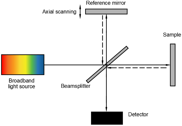
A schematic image of the OCT. Image Credit: Avantier Inc.
OCT Imaging Principles and Optical Requirements
OCT uses low-coherence interferometry, in which light reflected from internal microstructures is compared to a reference arm. This method enables depth-resolved imaging with axial resolutions ranging typically from 1-15 µm.
Key Optical Requirements:
Source: Avantier Inc.
| Parameter |
Typical Value |
Optical Impact |
| Axial resolution |
1–15 µm |
Requires broadband, low-coherence light |
| Lateral resolution |
5–25 µm |
Dictated by focusing optics NA and aberration |
| Optical path length diff. |
<1 µm across the system |
Requires wavefront-optimized optics |
| Chromatic dispersion |
Minimized or balanced between arms |
Requires careful material and design selection |
Key High-Precision Optical Components in OCT
1. Collimators and Beam Expanders
- Near-diffraction-limited quality and low divergence must be maintained (less than 1 mrad)
- Often made using aspheric doublets or molded glass optics to reduce spherical aberration
- Materials utilized: Fused silica, N-SF11, N-BK7, or IR-transparent glasses (for 1.3 µm OCT)
2. Scanning Optics (Galvo + MEMS Mirrors)
- Essential for producing 2D and 3D OCT volumes
- Requires angular precision (less than 0.01°), low inertia, and high-flatness mirrors (<λ/10)
- Reflective coatings optimized for 1060 nm, 1310 nm, or 1550 nm bands
3. Interferometric Beam Splitters and Couplers
- Employs polarization-maintaining (PM) fiber splitters or free-space plate beamsplitters
- Tolerances include transmitted wavefront error (less than λ/10) and surface flatness (less than λ/20)
- Dielectric coatings must maintain a 50:50 or custom ratio, with deviation less than 1 %
4. Focusing Optics (Scan Lens or Objective)
- Requires minimal aberration, whilst also delivering tight focus (high NA)
- Common types include achromatic doublets, GRIN lenses, or bespoke aspheres
- To ensure optimal spot quality, a threshold of below 0.05λ RMS Wavefront error should be met
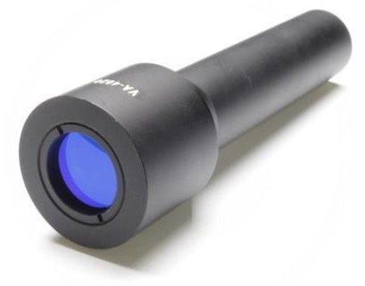
A beam expander. Image Credit: Avantier Inc.
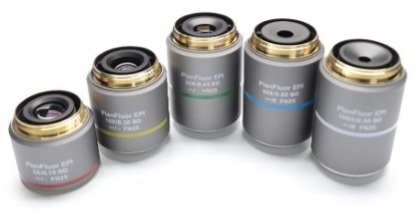
Objective lenses. Image Credit: Avantier Inc.
Benefits of High-Precision Optics in OCT
Source: Avantier Inc.
| Benefit |
Resulting Performance Gain |
| Lower wavefront error |
Enhanced image contrast and axial resolution |
| Reduced chromatic aberration |
Consistent focus across wide spectral bandwidth |
| High surface quality (e.g., 10-5) |
Minimal scattering and ghosting artifacts |
| Stable thermal and mechanical design |
Maintains alignment over time and environmental shifts |
Example: An OCT system designed with 0.01λ RMS optics can improve signal-to-noise ratio (SNR) by over 5 dB compared to a 0.1λ system - critical for imaging transparent or low-scattering tissues.
Fabrication Challenges
1. Tight Surface Figure and Roughness Requirements
- Challenge: Achieving a sub-λ/10 surface accuracy with a scratch-dig of 10-5 or better
- Solution: CNC polishing, magnetorheological finishing (MRF), and pioneering interferometry-based quality assurance
2. Broadband Dispersion Control
- Challenge: OCT systems often operate over bandwidths exceeding 100 nm, requiring optics that avoid chromatic mismatch
- Solution: Employ achromats, dispersion-compensated coatings, and matched material pairs
3. Compact, Integrated Packaging
- Challenge: Both Endoscopic & handheld OCT probes need miniaturized precision assemblies
- Solution: Utilize molded glass aspheres, monolithic lens barrels, and an alignment-free bonding approach
4. Coating Performance at NIR Wavelengths
- Challenge: Broadband AR coatings must maintain reflectance below 0.5 % across ranges like 800–1300 nm or 1000–1700 nm
- Solution: Use an electron beam evaporation coating
Representative Examples of OCT Systems and Optical Design
Example 1: Ultra-High-Resolution OCT for Retinal Imaging
System Type: Spectral-Domain OCT (SD-OCT)
Typical Wavelength: 840 nm ± 50 nm
Axial Resolution: Roughly 3 µm in tissue
Technical Highlights
- High-performance scan lenses devised to achieve diffraction-limited focusing (e.g., NA around 0.1, wavefront error less than 0.02λ RMS)
- Fused silica collimators with approximately 8 mm beam diameters and near-Gaussian beam quality (M2 < 1.05)
- Broadband beamsplitters optimized to decrease dispersion and reflection loss
Application Impact
Widely used in ophthalmology, OCT systems allow for the detailed examination of fine retinal structures. Precision optics enable cellular-level imaging of the retina, including photoreceptor mosaics and nerve fiber layers. This level of resolution supports early diagnosis of conditions such as glaucoma and age-related macular degeneration (AMD).
Example 2: Miniature Intravascular OCT Probes
System Type: Catheter-based Time-Domain OCT
Typical Wavelength: 1310 nm
Axial Resolution: Roughly 10 µm
Probe Diameter: Roughly 1.2 mm
Technical Highlights
- Bespoke GRIN lenses and microprism assemblies for compact light delivery
- Optical alignment held within ±5 µm eccentricity across the full 360° rotation
- Final optics typically have a surface roughness of less than 5 Å RMS and are AR-coated for broadband operation (1310 ± 100 nm)
Fabrication Considerations
- GRIN lens blanks can be fabricated utilizing precision glass molding
- Optical elements are aligned via active optical feedback systems and UV-cured adhesives
- Optical assemblies are often examined pre-integration utilizing fiber-coupled OCT interferometers
Application Impact
Miniaturized OCT probes are critical for intravascular imaging applications, supporting real-time imaging of coronary arteries with full circumferential coverage and shallow penetration depths. This enables early detection of plaque and vascular abnormalities.
Achieving Micron-Scale Resolution and Low-Noise OCT
High-precision optics are the pillar of OCT systems. With applications ranging from catheter probes for cardiovascular imaging to benchtop systems for retinal diagnostics, achieving micron-scale resolution and low-noise performance requires optics with excellent surface figures, minimal aberration, and consistent broadband performance.
Engineers can now produce miniaturized, robust, and highly accurate OCT optics, expanding the horizons of biomedical imaging and diagnostics, through the combination of state-of-the-art materials, manufacturing techniques, and optical design.
References and Further Reading:
- Wojtkowski, M., et al. (2004). Ultrahigh-resolution, high-speed, Fourier domain optical coherence tomography and methods for dispersion compensation. Optics Express, 12(11), p.2404. https://doi.org/10.1364/opex.12.002404.
- Drexler, W., et al. (2001). Ultrahigh-resolution ophthalmic optical coherence tomography. Nature Medicine, 7(4), pp.502–507. https://doi.org/10.1038/86589.
- Tearney, G.J., et al. (2012). Consensus Standards for Acquisition, Measurement, and Reporting of Intravascular Optical Coherence Tomography Studies. Journal of the American College of Cardiology, 59(12), pp.1058–1072. https://doi.org/10.1016/j.jacc.2011.09.079.
- Wang, L. V., Wu, H. I. (2012). Biomedical Optics: Principles and Imaging. (2024). (online) Wiley. Available at: https://www.wiley.com/en-us/Biomedical+Optics%3A+Principles+and+Imaging-p-9780470177013 (Accessed 21 Aug. 2025).

This information has been sourced, reviewed, and adapted from materials provided by Avantier Inc.
For more information on this source, please visit Avantier Inc.