Many modern bioscience techniques are based on applications of microscopy. To get the most satisfying results, it is important to know the fundamental basics of microscopy as well as the high-end methods. The Leica Science Lab is an interactive open access knowledge portal which wants to enlighten beginners and experienced practitioners and scientists about microscopy. To this end, a major focus of the Science Lab is basic principles, such as contrasting methods, Koehler illumination and fluorescence techniques or fluorescence itself. In addition, the Science Lab provides deep knowledge about current sophisticated microscopic applications and high-end techniques like CARS, TIRF or super-resolution microscopy.
A highlight of the Leica Science Lab is the way it presents the content: Information is packaged into scientific articles, white papers and tutorials with animated graphics which are assigned to several topics. To give you an idea about the sort of content which is available from the Science Lab platform, this article gives a short summary of one topic called "Basics in Microscopy".
From quantum states and Koehler Illumination
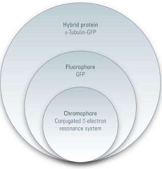 |
| Fig 1. Hierarchy in fluorescent proteins |
The "Basics in Microscopy" unit gives insights into fundamental theory and techniques in microscopy. It starts with several tutorials, including "Photo Effects of Light", "Basic principles of Luminescence" and "An Introduction to Fluorescence". In these articles, the reader gets in touch with the physical and chemical principles that enable us to use microscopy (mainly fluorescence microscopy) for varied scientific research. The tutorials explains how fluorescence works, and the characteristics of a fluorochrome are detailed. Electron quantum states, quenching, bleaching, and photostability are also discussed, and other effects like phosphorescence are explained.
Further articles focus on the technical background of microscopy. For example, optical contrast methods are explained.
Sometimes it is not possible to stain your specimen with the help of fluorescent agents. In this case, it is important to get the most information out of your sample with the help of optical tricks.
Simple brightfield microscopy is not sufficient to see small details. Therefore, scientists have invented different contrast methods to overcome that restriction. These methods try to change the phase shift, which is caused by the interaction of light with the specimen, into an amplitude shift. Its physical fundaments are explained in several tutorials like "Optical Contrast Methods - Physical Background and Fields of Application" or "Phase Contrast - Making Unstained Phase Objects Visible". In the case of phase contrast, amongst others, its exertion is perfect to make flat cells visible.
.jpg)
.jpg)
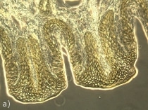
Fig 2. Cross section of rabbit taste-buds - one sample with three different contrast methods: (a) Phase contrast (b) Differential Interference Contrast (c) Bright field
Another way to enhance the contrast of an unstained specimen is to use Differential Interference Contrast (DIC). There is a great tutorial about this contrast method which is often used for imaging of living unstained or thick specimens, which employs an animation to demonstrate perfect illumination by DIC "Differential Interference Contrast - Step by Step Guide to Optimal DIC Setup".
When it comes to material science, a different contrast method is used for the identification of minerals, etc. By visualizing characteristic refraction properties and colors, polarization contrast enables the user to define a material. Even in biological scenarios, polarization contrast can be helpful. Crystals of cellulose can be identified by this method which is described in this tutorial: "Polarization Contrast - an Introduction".
A prerequisite for all of these contrast methods is perfect adjustment of the microscope. The best way to ensure optimal illumination is to use the Koehler principle. In the Leica Science Lab there is an easily understandable tutorial describing this standard procedure. With the help of animated instructions, "Koehler Illumination - Step by Step Guide to Optimal Illumination of Specimen" introduces the reader to the basic configuration of a microscope.
Microscopic pitfalls and how to avoid them
Once the mechanical setup is complete, the microscope user is able to watch his favorite cells, proteins etc. with his own eyes. But what about conserving these impressions? To make a record of your observations, it is necessary to have a powerful digital camera. A nice essay about making the right choice of camera is provided by "Digital Cameras - Beware of Pixel Mania". Here you can learn that it is not always best to have the camera with the most pixels...
Another article which aims to correct false assumptions in microscopy is "Beware of 'Empty' Magnification". Here, the difference between resolution and magnification is clarified, and their relative importance to microscopy is described.
Theoretical data about the achievement of a good picture - especially how depth of field is produced in microscopy - is provided by the article "How Sharp Images Are Formed - Depth of Field in Microscopy". Here, another clash between two parameters is described, which in theory are inversely correlated. These are depth of field and resolution.
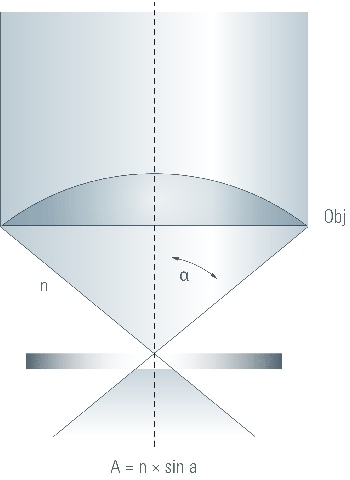 |
| Fig 3. The numerical aperture of the objective determines the detail resolution and brightness of the image. |
If you are worrying that the Leica Science Lab is all about blank physical parameters, never fear. A more less technical problem one has to consider is the microbiological environment on the surface of a microscope. Very often, microscopes are used by a lot of different people. All these users bring masses of germs with them. A way to avoid unnecessary pollution with microorganisms is to cover instruments with a silver coating. "Antimicrobial Coating for Educational Microscopes: A Contribution to Laboratory Hygiene" talks about that advancement and gives explanations why this makes sense for the user.
Learn. Share. Contribute
To conclude, the Leica Science Lab wants to inform the user about all the aspects of microscopy, and the opportunities it provides, from the fundamentals to high-end applications. In case of the topic "Basics in Microscopy" which is advertised here, the user is introduced to essential optical problems and is confronted with the proper usage of a microscope bit by bit. Furthermore, the operator can read about the professional presentation of the acquired data. In every case, the aim is to involve the user in the learning process by integrating them into the Science Lab community - either by discussing things in the "community box" or by contributing with their own article. This will help to install the Leica Science Lab as a lively scientific interaction platform that supports the researcher in his everyday work. Come and join us!
To increase the amount of valuable content in the resource, Leica Science Lab invites YOU to be one of the growing community of Science Lab authors. The number of authors has reached 150 and is still growing steadily. As an author you can participate in discussions about others' articles, as well as contributing your own material.

This information has been sourced, reviewed and adapted from materials provided by Leica Microsystems.
For more information on this source, please visit Leica Microsystems.