Tabletop SEMs have undergone significant advancements over the past decade, positioning themselves as the ideal choice for users seeking to go beyond the capabilities of optical stereo microscopy.
These devices allow for the inspection of objects at higher magnification and resolution, surpassing the limitations of traditional optical microscopy, such as a shallow depth of focus.
In addition, the backscattered electron detectors of typical tabletop SEMs provide extra insights into the distribution of different materials, which can be quantified by using proper EDX analysis techniques.
The technical implications of instrument miniaturization and the restrictions imposed by the cost of tabletop SEM concept place natural constraints on the obtainable image resolution. Users who frequently need to resolve structures smaller than about half a micrometer may not be satisfied with a tabletop SEM.
Previously, users with an increased imaging demand had no choice but to invest in a traditional room-filling SEM with a large footprint, including external infrastructure, and an unhindered operation concept that typically required expert knowledge to fully utilize the instrument.
This article proposes a smart solution to this challenge: the Hitachi FlexSEM1000, an incredibly powerful little SEM with a new compact layout designed to address the abovementioned issues.
It combines a high and easily achievable performance comparable to a traditional full-scale SEM with an ultra-compact (just 45 cm wide) tabletop-SEM-like body, user-customizable software, entirely user-maintainable, and extremely appealing price tag.
Instrument Concept and Outline
A common issue with a typical ultra-compact tabletop SEM is that its compact size comes at the expense of functionality: The vacuum system is limited to a specific operation mode, and the diminutive electron optic system prevents the use of high-quality low-aberration lens systems.
This can be partially compensated for by employing special electron sources, such as LaB6 / CeB6 or even Field Emission sources, which can enhance the resolution of compromised electron optics, but such approaches usually cause more problems than advantages when it comes to general functionality, reliability, handling, and cost.
This was not the direction Hitachi wanted to take while creating a high-performance yet extremely small and mobile new SEM.
FlexSEM in stand-alone setup. Image Credit: Hitachi High-Technologies - Europe
FlexSEM in desktop setup. Image Credit: Hitachi High-Technologies - Europe
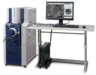
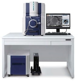
The FlexSEM is compact, weighing only 165 kg, and very mobile, thanks to its wheeled chassis. It can be installed on a workstation (similar to a tabletop system) or used on its own, and it requires no external facilities except for a 230 V power outlet and a tiny rotary pump.
Nevertheless, it is still sufficiently sized to accommodate traditional high-performance Tungsten-source-equipped electron optics with up to 20 kV accelerating voltage, as well as a sophisticated TMP-driven vacuum system that enables both a proper high-vacuum mode for high-resolution imaging and a dedicated and manageable low-vacuum mode with adjustable chamber pressures up to 100 Pa.
This allows for the observation and examination of uncoated non-conductive materials without charge-up and, when combined with an optional Peltier cooling stage, the observation of damp biological samples.
The 5-axis specimen stage, which can tilt from -20° to +90°, can be entirely withdrawn from the chamber to allow for the easy placement of samples up to 80 mm in diameter and 40 mm in height. An EDX system and a chamber access port can also be installed to connect other devices.
When not in use, the entire system can be completely shut down and restarted in less than five minutes.
Operation complexity may be easily changed to meet the skills and demands of each user, and all operations are supported by sophisticated automated functions, such as a color specimen camera for sample stage navigation, a stage trackball, recipe memory, an operating knob panel, and so on.
The Heart of the FlexSEM: Electron Optics
The FlexSEM1000 is built on powerful electron optics that cover an accelerating voltage range of 300 V up to 20 kV, ensuring an SE image resolution of 4 nm at high vacuum settings.
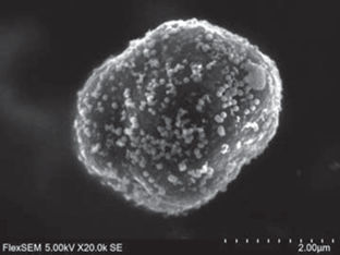
Figure 2a. The toner particle was imaged at 5 kV in high-vacuum mode with the Everhart-Thornley-SE detector. Magnification (polaroid base) is 20.000x. Image Credit: Hitachi High-Technologies - Europe
While 20 kV is usually needed and is adequate for carrying out EDX elemental identification of most elements in compliance with ISO standards (for example, asbestos analysis), such high accelerating voltages are rarely optimal for best real-world sample imaging when fine surface structure details are desired.
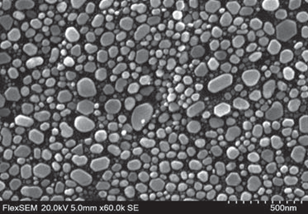
Figure 2b. Gold grains on a carbon substrate. Accelerating voltage 20 kV, magnification 60.000x. Image Credit: Hitachi High-Technologies - Europe
The suggested working settings range from low to medium, accelerating from 1 kV to 10 kV. The FlexSEM1000 meets such requirements: its dynamic gun bias system ensures a nearly constant high gun brightness across the accelerating voltage range.
A double-condenser-optics system and a low-aberration objective lens shape this bright electron beam into a small spot on the sample, allowing for high-quality imaging with an SE resolution of just 15 nm at 1 kV of accelerating voltage.
Adjusting the excitation of the condenser lens, one can precisely change the probe current over a broad range, whereas condenser lens two continually optimizes beam admission into the objective lens.
For ease of use, the user must choose a “spot intensity” number between 1 and 10; there is no need to worry about such lens details. The FlexSEM1000 has no movable apertures therefore beam alignment is automated and highly stable over time.
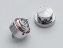
Image Credit: Hitachi High-Technologies - Europe
The FlexSEM1000 generates electrons using pre-centered cartridge-type tungsten hairpin filaments. They are “ready to use”; when a filament exchange is needed, the FlexSEM specimen chamber is ventilated, the gun is turned open, and the old filament with the Wehnelt suppressor screwed on is removed.
The Wehnelt cap is attached to the new filament cartridge placed in the gun holder.
The gun head is flipped back, the chamber is evacuated, and a single function in the “Beam Adjustment” menu box completes the process: Saturate the filament heating current and adjust the beam path via the optics for maximal beam current on the sample.
The entire process takes less than 10 minutes and may be completed by any user (with optional step-by-step menu instructions).
With this approach, tungsten filaments are the perfect electron sources for a powerful yet compact SEM like the FlexSEM1000 because of their general robustness, low vacuum needs, and, most importantly, low cost.
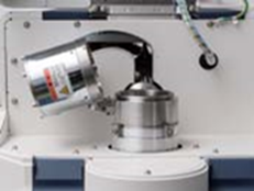
Figure 3. FlexSEM’s electron gun flipped open for direct access to the filament cartridge. Image Credit: Hitachi High-Technologies - Europe
The Detection System of the FlexSEM
The FlexSEM1000 includes two specialized vacuum operation modes: a high-vacuum mode for observing electrically conductive samples and a variable-pressure mode for analyzing non-conductive or damp samples with chamber pressures of up to 100 Pa.
As a result, the FlexSEM comes pre-installed with appropriate electron detectors. In addition to an Everhart-Thornley-type detection system for high-resolution SE imaging in high-vacuum mode, five high-sensitivity backscattered electron detectors positioned on a carrier arm below the objective lens can be used in both vacuum modes.
Figure 4 depicts the arrangement of the BSE detector set: Four detectors are positioned as 90° segments of a circle, with the fifth element located to the side.
This geometry enables the creation of various image impressions: When the circle segments are combined, a uniformly lit material/crystal orientation contrast-rich image is obtained (“BSE-COMPO”).
When the fifth (side) detector is added to the circle, a variable topographical (shadow) impression is added to the “conventional” material /orientation contrast image.
The result is a three-dimensional impression (“BSE-3D”), enabling the judgment of the specimen’s shape and the material/crystal data. Pure topographical images can be obtained by grouping two neighboring detectors and removing their signal (“BSE-TOPO”).
The optional 3D-viewing capability allows for the simultaneous readout of four quadrant detector images. The Hitachi Map3D software package converts these four directional images into a quantitative surface height image of the sample, from which metrics such as roughness, step heights, etc., can be retrieved.
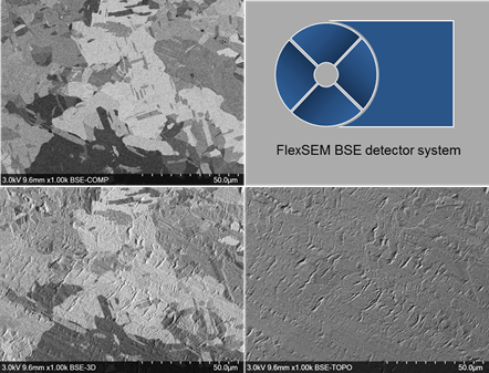
Figure 4. Examples of the different BSE imaging modes can be seen on the example of a rolled and briefly Ar ion beam polished Cu foil. At the upper-left, only grain orientation contrast without any topography is seen by the COMPO mode. At the lower left, by the 3D setup, sample topography (rolling traces) appear overlaid on the grain orientation contrast. At the lower-right, pure topographical information is displayed by the TOPO mode. By changing the segment grouping the “illumination direction” can be adjusted in 90° steps – this helps to emphasize or suppress pattern running preferentially in certain directions. Image Credit: Hitachi High-Technologies - Europe
The FlexSEM BSE detector system can operate efficiently from low accelerating voltages of about 1.5 kV to a maximum accelerating voltage of 20 kV.
This enables it to carry out very surface-sensitive yet multi-mode BSE imaging on even sensitive or very finely structured samples and give high-contrast images under the conditions required for EDX analysis. In short, it is an ideal detection device for all situations, from high to low vacuum and from low to high accelerating voltages.
The BSE detection system is the default image sensor in the low-vacuum mode, where the traditional Everhart-Thornley SE detector cannot be used. With its variable imaging modes, it can create highly topographical images in addition to the “flat” material contrast images, making it suitable for most applications.
Some samples may still need “true” SE imaging in the low-vacuum mode. Usually, these are organic (low atomic weight) materials with a low BSE yield or high-aspect-ratio objects that must be scanned across long working distances.
Backscattered electrons are absorbed by interactions with gas molecules in the chamber, causing BSE imaging to become weak (blurry) over long working distances and at high chamber pressures.
The optional low-vacuum SE detector “UVD” can help in this instance. This detector technique converts secondary electrons generated from a specimen into optical photons that may travel the distance between the specimen and the detector with minimal chamber gas interactions.
Unlike previous-generation low-vacuum SE detectors, which rely on charge cloud drift from specimen to detector electrode, the UVD produces contrast-rich SE-like images that are relatively independent of working distance and low-vacuum-mode chamber pressure.
Figure 5 depicts an example of a superabsorbent polymer photographed with the UVD at 3 KV accelerating voltage and 30 Pa chamber pressure.
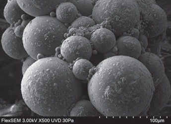
Figure 5. Super-absorbent polymer. No metal coating applied. Low-vacuum mode 30 Pa, SE signal (UVD). Image Credit: Hitachi High-Technologies - Europe
Because of its “optical photon counting” approach, the UVD can function as a monochrome cathode-luminescence (CL) detector in high-vacuum mode. In addition, FlexSEM’s multi-detection technology allows for the concurrent display and recording of two signals in dual full-screen mode.
Operability and User Interface
The FlexSEM adapts to each operator’s skill level. The system’s fundamental notion is “convenient & risk-free operation”.
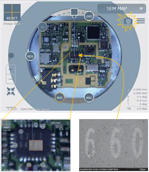
Image Credit: Hitachi High-Technologies - Europe
During sample loading, precautions are taken to avoid collisions between the sample and the SEM lens or detectors. Cup-like sample containers with height-adjustable inner stubs make it simple to select the proper sample height.
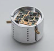
Image Credit: Hitachi High-Technologies - Europe
Simply put, no specimen component may extend beyond the upper edges of the cup-shaped holder.
This is verified when the drawer-type chamber door is closed. A height gauge at the chamber entrance, similar to the objective lens and the BSE detector, contours inside the specimen chamber and prevents any sample of inappropriate height from entering the SEM chamber (skilled users are free to use conventional sample holders).
When tilting the specimen stage during examination, the FlexSEM software automatically determines safe limits for tilt angles and operating distances based on the sample size. Although the FlexSEM’s Tilt and Z axes are not motorized, the FlexSEM software monitors them to ensure the current status is always known.
The FlexSEM includes a color camera at the specimen chamber door to aid orientation and stage navigation. This camera takes a high-resolution color image of any loaded specimen and displays the image in the “SemMap” navigation window.
The image can be zoomed in for better orientation, rotated with stage or scan direction changes, and the position of important detectors is noted at the image’s periphery for a clear comprehension of any directional or shading effect.
It is also feasible to import externally created images and evaluate them with a 3-point-alignment to facilitate navigation.
Ultimately, a Snapshot button allows the instrument to rapidly patch the currently displayed low-magnification SEM images right into the appropriate spot of the optical navigation image, enabling locally fine navigation based on the definition of the SEM image.
For reporting reasons, the navigation image can be exported with the positions of recent SEM image acquisitions on the specimen marked on it; the marking numbers correspond to the image numbering when saved in auto-naming mode.
The safe approach for specimen registration, photo documentation, and loading is the same for all users. However, three different skill levels can be used for actual SEM operations.
For absolute beginners who may be unfamiliar with SEM-typical terminology, such as “accelerating voltage” or “spot intensity”, and how to choose the right one for the desired application, the FlexSEM provides a list of “application purposes”.
These are basic requirements for each SEM investigation, such as “surface composition”, “surface topography”, or “EDX analysis”, among others.
For each application, the FlexSEM1000 has a pre-defined and self-aligned optimal working condition known to produce the appropriate output for the user. This allows even inexperienced users to quickly capture good-quality images of their sample in a short time.
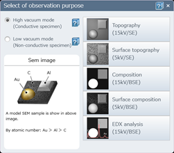
Image Credit: Hitachi High-Technologies - Europe
For common tasks and relatively experienced users, the FlexSEM includes an “operating condition” recipe memory.
FlexSEM super users can retain individually set conditions that have been proven to be optimal for frequently necessary (repeating) tasks. Choosing a recipe loads the vacuum mode, the electron optical conditions, and the detector assignment on the primary and secondary screens.
For skilled users, FlexSEM provides complete access to all electron optical parameters within the allowable limits.
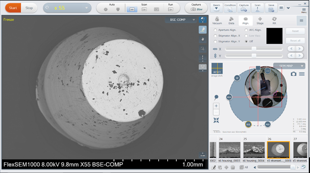
Image Credit: Hitachi High-Technologies - Europe
The FlexSEM1000 can be used in several ways. All functions are standard and can be operated using a PC mouse or a touch screen. There are automated brightness/contrast and focus capabilities that are both reliable and speedy.
The instrument can be connected to a trackball for manual stage X and Y fine operation, as well as a “knob panel”, including the most commonly used functions, such as focus, brightness/contrast, electrical image fine shift, scan speed settings, and magnification, among others.
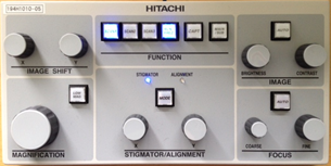
Image Credit: Hitachi High-Technologies - Europe
This knob panel is extremely popular among experienced SEM users, and it can provide higher operation throughput than a mouse-only operating style. The “quick access button” option can be customized for each login profile.
While a novice user may prefer a clean user interface with the bare minimum of buttons and functions to avoid being distracted by a large number of “unknown” function buttons, a more experienced user may prefer quick access to useful and frequently used functions without having to navigate the main menu tree. The FlexSEM1000 is versatile enough to accommodate all these options.
Advanced Analytics: EDX, 3D Metrology, Particle and Fiber Metrology
The FlexSEM1000 also offers specialized analytical applications that extend beyond simple imaging.
For instance, it supports professional elemental EDX analysis tools from prominent firms like Oxford Instruments, Bruker, and EDAX. Unique tiny detectors with 30 mm2 SDD can be integrated entirely into the FlexSEM1000’s body, enabling high count rates of several kilo-counts per second.
The instrument’s maximum accelerating voltage of 20 kV enables K-line detection of higher atomic weight elements and meets DIN/ISO standards for asbestos analysis. Point/area spectra, line scans, and element distribution mappings are all available.
Use is simple: load an appropriate operating condition from FlexSEM’s recipe memory, then run the EDX program on a regular Windows 10 PC. Images and EDX results can be saved to the same data folder.
Another benefit of adopting EDX expert equipment is the ability to upgrade the EDX software to a professional level, including automated routines like automatic particle analysis.
Another unique analytical approach enabled by the equipment is elevating traditional 2-D SEM to the third dimension. The four quadrants of the BSE detector circle can produce four images of the same object simultaneously, but they are “quasi-illuminated” from each detector element’s perspective.
The optional Hitachi Map3D Software, when combined with a unique height calibration method, reconstructs an accurate 3D surface model of the inspected sample region.
Height profiles can be retrieved to determine step heights and surface roughness metrics following ISO standards. Surface and volume measurements are also feasible.
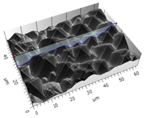
Image Credit: Hitachi High-Technologies - Europe
Hitachi Map3D has several additional useful capabilities, including pore/particle extraction and metrology, stitching of large high-pixel-density image fields from individual images recorded with the optional “ZigZag-capture” module, and basic image segmentation for colorization.
Maintainability and Cost Considerations
For periodic preventive care of the FlexSEM1000, the user has the option of using a professional Hitachi service or a complete do-it-yourself approach.
The tungsten hairpin filament cartridge-type electron source is simple to change and activate within minutes. All fixed electron optics apertures are positioned within a “liner tube” that can be withdrawn entirely from the SEM column for aperture changing and cleaning.
This is especially helpful in settings where a charged particle or fiber has migrated to an aperture: Simply unlock the gun head, pull out the liner tube, examine and eventually clean/replace the aperture, and re-insert it. There is no need to hire a service specialist or pump the system for 24 or 60 hours before using it again.
The FlexSEM1000's annual maintenance cost is less than €1,000, which is a significant consideration when choosing a compact SEM.
Conclusion
The Hitachi FlexSEM1000 is a small, powerful, and easy-to-use SEM. Its performance and functionality range are identical to today’s full-scale variable pressure SEMs and significantly exceed the capabilities of traditional tabletop SEMs. It may adapt to a user’s demands and talents.
In addition, its minimal maintenance expenses and reasonable price make it an appealing option.

This information has been sourced, reviewed and adapted from materials provided by Hitachi High-Technologies - Europe.
For more information on this source, please visit Hitachi High-Technologies - Europe.