Infrared-based chemical imaging has been a powerful analytical technique for decades. It delivers spatially resolved information about the chemical composition of a sample, label-free and non-destructive. For pathologists studying tissue, pharmaceutical scientists analyzing tablets, and environmental researchers tracking microplastics, it is an indispensable tool.
The Problem: Measurement Time as a Bottleneck
The technique, however, was held back from widespread routine due to its lack of speed speed. Conventional FT-IR imaging systems require hours to days for larger sample areas. In clinical research, pharmaceutical quality control, or particle analysis, this forces a difficult trade-off - either accept long wait times or sacrifice spatial resolution and spectral quality. This dilemma has delayed the translation of promising IR imaging methods into clinical and industrial practice for years.
But the solution, is readily available: IR Laser Imaging with the LUMOS II ILIM.
Meet the LUMOS II ILIM - the next generation of IR Laser Imaging
There's a Paradigm Shift in IR Microscopy
From Spectra to Images
With the LUMOS II ILIM, Bruker has integrated a fundamentally new approach into its established LUMOS platform: Infrared Laser Imaging (ILIM). Rather than scanning point by point, the system captures complete chemical images in a fraction of the time previously required. A published study in the Analyst on TMA (Tissue Microarray) cores puts this into numbers.
| Parameter |
FT-IR |
ILIM |
| Imaging Speed |
~370 spectra/s |
62,400 spectra/s |
| Field of View |
<0.5 mm² |
2.2 x 2.0 mm² |
| Full TMA (1,500 cores) |
~3 months |
~2 days |
| Area Scanning Speed |
Baseline |
Up to 169x faster |
These speed gains do not come at the expense of data quality. In fact, the higher intensity of the laser source allows creation of IR images that are much sharper. A patented spatial coherence reduction technology eliminates typical laser artifacts, producing chemical images with a clarity and fidelity that were not achievable with conventional systems.
3 Months Compressed to 2 Days by ILIM
What these numbers mean in practice is best illustrated by the experience of Prof. Peter Gardner at the University of Manchester, who has worked in tissue imaging and spectral pathology for over two decades:
We have been working in the field of tissue imaging and spectral pathology for over 20 years. One of the key barriers to clinical adoption is the long measurement times for full hyperspectral imaging of large areas of tissue. A sample set consisting of nearly 1500 prostate tissue cores, that took 3 months to measure on our old instrument, was measured in just two days on the Bruker ILIM system. This is a game changer!
Peter Gardner, Ph.D., Professor of Analytical and Biomedical Spectroscopy, University of Manchester
Gardner's study confirms something important: A researcher who previously had to choose between statistical power and measurement time no longer has to make that trade-off with IR laser imaging.
Start by Discovering the ILIM Applications
TissuePlusTM
Image full tissue microtome sections in minutes, not hours, giving you statistically meaningful datasets within clinical timelines. Pair it with MALDI imaging and the infrared image acts as a map for targeted mass spectrometry, cutting acquisition time dramatically without losing molecular detail.
Learn more about TissuePlusTM here.
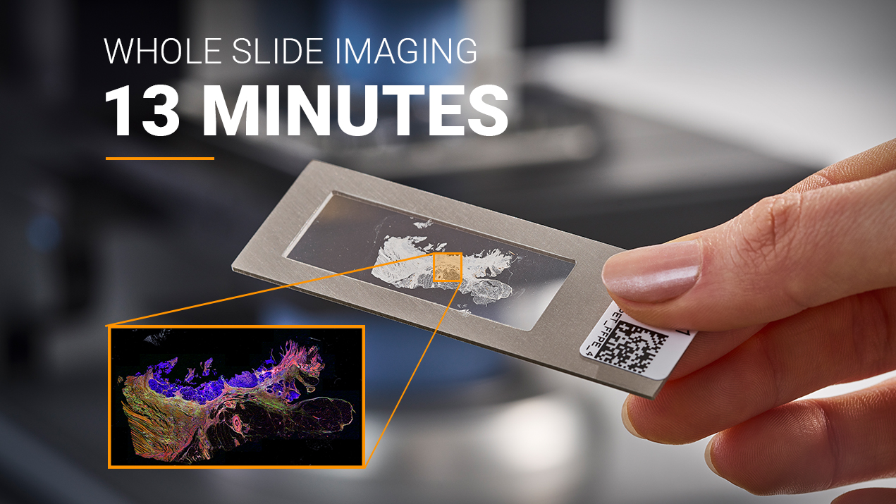
Image Credit: Bruker Optics
TabletPlusTM
Chemically map active ingredients, excipients, and fillers across tablets in minutes. For pharma QC and deformulation, this is a major step up from classical methods, with faster release testing and shorter formulation development cycles.
Learn more about TabletPlusTM here.
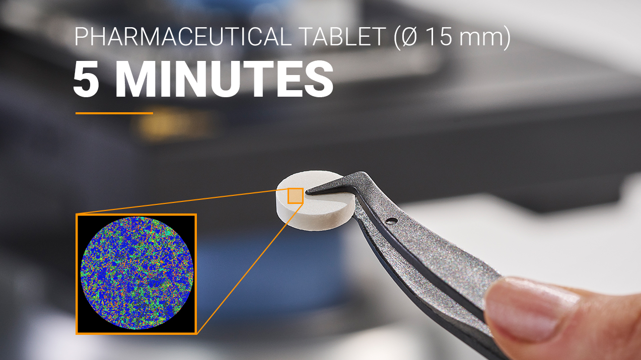
Image Credit: Bruker Optics
ParticlePlusTM
Analyze complete filters in minutes, with chemical ID down to individual particles. As microplastics regulations tighten around drinking water and environmental compliance, that throughput matters, especially for labs handling high sample volumes.
Learn more about ParticlePlusTM here.
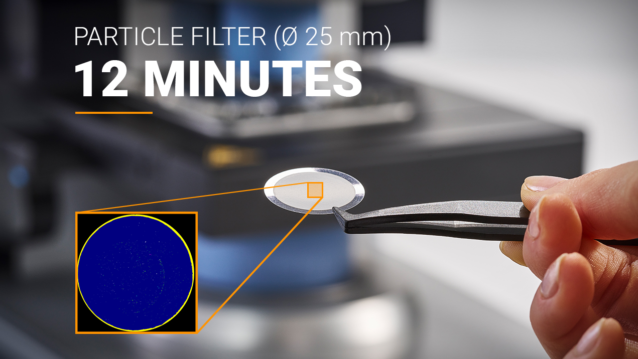
Image Credit: Bruker Optics
The Next Step? See it in Action!
Bruker offers live demonstrations and application consultations