May 6 2014
Confocal microscopy is a versatile imaging method, through which viewing cells and tissues is made possible. It is a major improvement from conventional light microscopy, as it also helps create images in 3D.
The term ‘confocal’ is used when two lenses are arranged to focus on the same point. The key optical difference between conventional and confocal microscopy is the presence of confocal pinholes in the confocal microscope, to allow only light from the plane of focus to reach the detector.
The two main types of confocal microscopes are the laser scanning microscopes, and the tandem scanning microscopes (TSM). The former is ideal for immunofluorescence microscopy, and the latter is used for high-speed reflection imaging.
Benefits of Confocal Microscopy
Confocal microscopy produces images with impressive resolutions of up to 1.4 times greater than that of conventional microscopy.
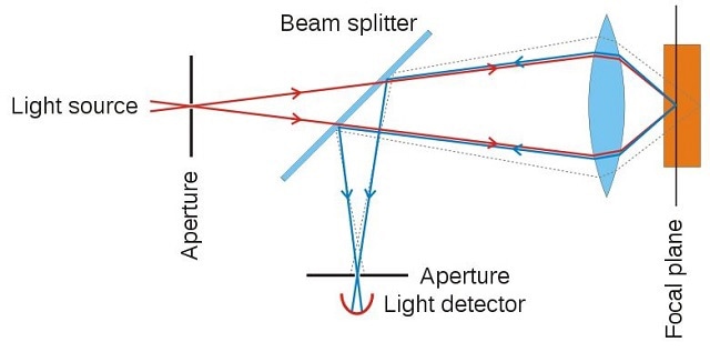
(Image source: Wikimedia Commons)
This is possible because it can eliminate out-of-focus light, using spatial filtering. The spatial filtering feature also helps to eliminate flare in samples that are thicker than the plane of focus. Computer software can be used to digitally reconstruct 3D illustrations of the sample. It offers relatively better resolutions, both vertically and horizontally, at about 0.5 and 0.2 µm, respectively.
These microscopes have a higher level of sensitivity, due to the in-built highly sensitive light detectors. They possess the ability to collect images captured over a period of time.
High-power laser illumination and reduction in light-scattering objects enables a less invasive form of imaging. This technology also allows non-invasive imaging of thick sections of semi-transparent tissues. It can be operated with relative ease.
Compared to conventional optical microscopy, this technology has a controllable depth of field, and the ability to gather serial optical sections from thick samples.
Laser-scanning confocal types can cut exceptionally clean, thin optical sections of about 0.5 to 1.5 µm out of thick samples measuring more than 50 µm, using either reflection or fluorescence. It can view samples in planes running parallel to the line of sight.
Other benefits include:
- Enhanced signal-to-noise ratio
- Comprehensible and close examination of thick samples
- Z-axis scanning and depth perception in Z-sectioned images
- Electronically adjusted magnification
Applications of Confocal Microscopy
In recent years confocal microscopy has been adapted into numerous fields - such as cell biology, life sciences, semiconductor inspection, materials science, neuroanatomy, and neurophysiology.
Key applications areas include:
- Stem cell research
- Photobleaching studies
- Fluorescence resonance energy transfer (FRET)
- Fluorescence recovery after photo-bleaching
- Fluorescence in-situ hybridization
- Lifetime imaging
- Multiphoton microscopy
- Total internal reflection fluorescence microscopy (TIRFM)
- DNA hybridization
- Membrane and ion probes
- Bioluminescent proteins
- Epitope tagging
Conclusion
Confocal microscopy is considered as a bridge between conventional widefield microscopy and advanced transmission electron microscopy. Due to the vast improvements in this technology, and the wide range of its applications in areas of recent research interest, confocal microscopy will likely continue to be well received in the scientific world.
References