Image Credit: D. Kucharski K. Kucharska/Shutterstock.com
In February 2020, a team of engineers reported the development of a technique that was able to enhance dark-field microscopy using a small, mirrored chip to generate images in place of the expensive components that were previously required by conventional methods. The advancement of the dark-field microscopy technique opened up the method for use in a variety of new applications, such as diagnostics and other bioanalytical applications.
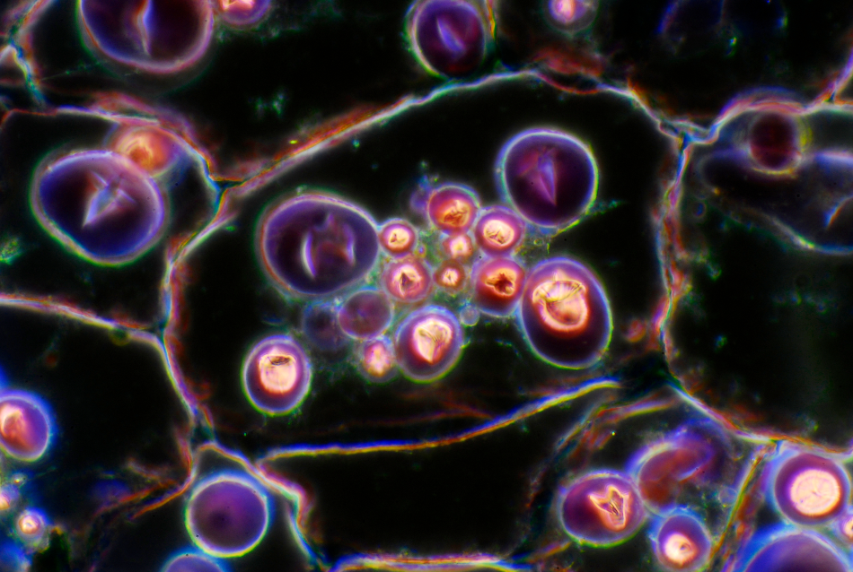
Dark-Field Microscopy Images Microorganisms in Impressive Detail
The method of dark-field microscopy has been used for many years by the biological sector to produce high-contrast images of a variety of subject matters, particularly of microorganisms in water, such as bacteria and algae, but also of blood cells and other microscopic biological structures. The images produced by dark-field microscopy can uncover the fine details of a sample that a regular bright-field microscope would fail to visualize in high focus and clarity.
The technique has numerous benefits that make it useful for imaging tiny biological systems. For example, it does not require sample staining, and it can accentuate small features that may be key to gaining an understanding of a sample.
The conventional method of dark-field microscopy causes light to scatter as it hits a sample by blocking off the light source. Usually, in coming into contact with an object, light scatters in all azimuths or directions. The dark-field microscope removes the light of the zeroth order, the dispersed light, resulting in just the scattered beams hitting the sample. A condenser is used to cause these rays to approach the sample at different angles instead of as a direct light source that hits the sample from above, as is the case with regular light-microscopy.
For this reason, dark-field microscopy can generate high contrast images of objects that appear bright against their dark background where light-microscopy may have failed to visualize the details of the sample where the refractive values may be similar to the background.
However, there are drawbacks to the traditional method of dark-field microscopy. The main disadvantage of the method is that it requires specialized components that are bulky and expensive, like objectives and condensers, for example.
Exploring Structural Colors Leads to Innovative Light-Emitting Chip
A team of engineers at the Massachusetts Institute of Technology (MIT) aimed to enhance the method of dark-field microscopy in order to overcome this limitation. In a paper published in the journal Nature Photonics, the engineers describe how they established a method of simplifying the equipment used in dark-field microscopy and enabled it to be manufactured as a much smaller device, opening up new potential applications of the method.
The lab had previously been focused on developing devices with "structural colors," those that are not reliant on dyes or pigments but are constructed of nanostructures that scatter and reflect light similarly to the way a prism does. Nature holds many examples of structural colors, such as the wings of a butterfly, or fish scales. The scientists at MIT used these natural examples as inspiration, guiding their exploration into how light can be manipulated from a microscopic and structural perspective.
The design and development of a tiny, three-layered chip were born out of this research, with it being initially intended to be used as a miniature laser. The chip's middle layer is constructed of tiny nanoparticles and acts as a light source when excited by fluorescent light. This occurs because a small amount of blue light has the effect of causing the quantum dots within the chip to emit bright orange and red colors, and, because of the stability of the dots, they can emit color for a long time.
A Bragg mirror was placed over the chip's light-emitting layer. This mirror is constructed of alternating nanoscale layers of transparent materials that have different refractive indices, resulting in differences in the amount of light reflected by the individual layers.
The arrangement of the layers in the Bragg mirror dictates how the photons escape out of the chip, only allowing them out when the light enters the chip at high angles. Underneath this light-generating layer, the researchers added a layer made out of a solid epoxy coated with a reflective gold film to capture and recycles the photons that the Bragg mirror rejects by bouncing it back up, where it enters the chip at a new angle.
A Hand-Held Dark-Field Microscope
The result of the work by the team at MIT saw a small, mirrored chip developed with the capacity to generate dark-field images without the need for bulky and expensive components. This chip is a little larger than the size of a postage stamp and as thin as a credit card was designed to be placed on the microscope's stage, emitting a hollow cone of light to create the highly detailed images of microorganisms, such as bacteria and algae, as well as other tiny translucent objects.
It is possible that the newly developed chip may be used in hand-held microscopes to make dark-field imaging portable, allowing it to be used in a wider array of applications.
References and Further Reading
Chazot, C., Nagelberg, S., Rowlands, C., Scherer, M., Coropceanu, I., Broderick, K., Kim, Y., Bawendi, M., So, P. and Kolle, M. (2020). Luminescent surfaces with tailored angular emission for compact dark-field imaging devices. Nature Photonics. https://www.nature.com/articles/s41566-020-0593-1
Rosman, C., Pierrat, S., Henkel, A., Tarantola, M., Schneider, D., Sunnick, E., Janshoff, A., and Sönnichsen, C. (2012). A New Approach to Assess Gold Nanoparticle Uptake by Mammalian Cells: Combining Optical Dark-Field and Transmission Electron Microscopy. Small, 8(23), pp.3683-3690. https://onlinelibrary.wiley.com/doi/abs/10.1002/smll.201200853
Taylor, M., and Bowen, W. (2013). Enhanced sensitivity in dark-field microscopy by optimizing the illumination angle. Applied Optics, 52(23), p.5718. https://www.osapublishing.org/ao/abstract.cfm?uri=ao-52-23-5718
Disclaimer: The views expressed here are those of the author expressed in their private capacity and do not necessarily represent the views of AZoM.com Limited T/A AZoNetwork the owner and operator of this website. This disclaimer forms part of the Terms and conditions of use of this website.