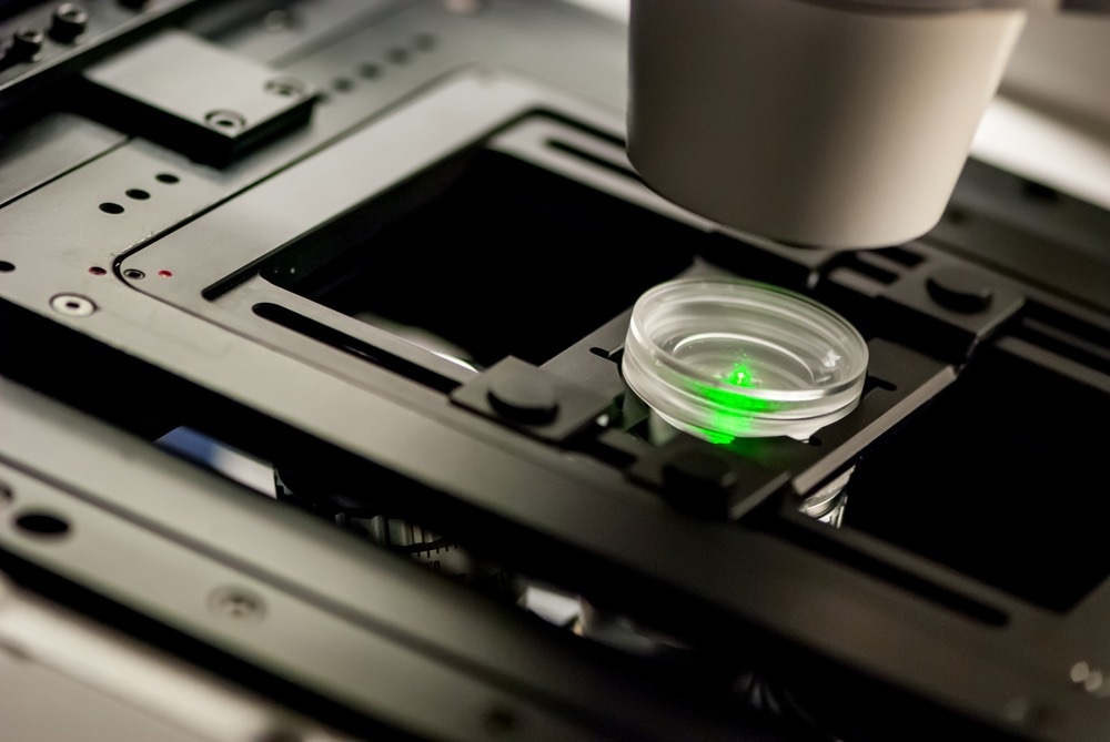
Although transmitted light microscopy techniques, including differential interference contrast (DIC), phase contrast, and polarized microscopy have improved the visualization of living specimens by enhancing their intrinsic contrast, live imaging using fluorescence microscopy has allowed life science enthusiasts to visualize subcellular structures at higher resolution.

Image Credit: Micha Weber/Shutterstock.com
The idea of studying biological materials using fluorescence was conceived in the early 19th century. However, its practical application was not demonstrated until 1938, when Richard Manly and Robert Goldstein first developed the fluorescent microscope.
What is a Fluorescent Microscope?
A fluorescent microscope is a powerful tool that selectively reveals the object of interest (signal) on a black background. Various fluorescent indicators, also called fluorescent probes or fluorophores, are tailor-modified to label specific targets such as lipids, proteins, or ions.
Fluorescence-labeled biomolecules stand out against a black background under a fluorescence microscope, providing an understanding of the role of various biomolecules in cell physiology and disease development.
How does a Fluorescent Microscope Work?
To understand the workings of a fluorescent microscope, it is important to become familiar with the underlying phenomenon of fluorescence. When light of a specific wavelength is irradiated on a fluorophore-labeled specimen, the electrons in the specimen are excited to higher energy levels. The subsequent de-excitation of electrons to attain stability causes the emission of light with longer wavelengths, which is called fluorescence.
However, the spectral emission during this phenomenon also includes weaker fluorescence, which is separated using a spectral emission filter to ensure the distribution of a single wavelength of light during the imaging process. Because the color of the light depends on its wavelength, the emitted light has a different color than the absorbed light.
Conventional Versus Fluorescent Microscope
While a conventional microscope uses visible light (400-700 nm) as the light source to irradiate the sample, a fluorescent microscope uses higher-intensity light to excite the fluorophores in the specimen. Furthermore, fluorophores emit light of longer wavelengths during fluorescence, producing a magnified image with a color different from that of the original light source.
Types and Applications of Fluorescent Microscopes
Given below are the various types of fluorescence microscopes and their principles and applications:
- Epifluorescence microscope: In this microscope, the illuminating light penetrates the full depth of the specimen, which facilitates the imaging of intense signals and co-localization studies on a sample. Images obtained using this type of microscope help study the molecular interactions and dynamics of gene expression.
- Confocal laser scanning microscope: This microscope increases the optical resolution and contrast in the image by eliminating out-of-focus light. This type of microscope is compatible with the three-dimensional (3D) imaging of molecular and cellular dynamics.
- Two-photon microscopes: This type of fluorescence microscope depends on the two-photon absorption principle, allowing visualization of living tissue at greater depths, which is otherwise unattainable using conventional or confocal microscopes. Hence, two-photon microscopy has been applied in cancer, embryonic, kidney, and neuroscience research.
- Super-resolution microscopes: These microscopes achieve high resolution by utilizing techniques such as stimulated emission depletion (STED) or photoactivated localization microscopy (PALM), which improves spatial resolution by circumventing the diffraction limit. A super-resolution microscope helps study the fine details of various biological materials that are not achievable using traditional fluorescence microscopes.
- Multiphoton microscopes: As evidenced by their name, multiphoton microscopes use lasers to simultaneously excite multiple light photons, to study the sample’s fluorescence in 3D space. This type of microscope is used to image intact biological tissues at a molecular level.
Advantages and Limitations of Fluorescent Microscopes
Fluorescent microscopes have several advantages, including high sensitivity, high resolution, multiplexing ability to simultaneously visualize multiple pathways in a sample, high specificity, non-destructive analysis of samples, ability to image live cells, and ability to trace the exact location of proteins.
However, there are limitations in fluorescence microscopy, including photobleaching of fluorophores due to chemical damage during fluorescence, phototoxicity of cells due to the generation of reactive species, excitation and emission overlap, leading to false results, limited sample thickness, limited multiple labeling, and observation of structures that are only fluorescence-labeled.
Latest Trends in Fluorescent Microscopy
A recent article published in the journal Nature Communications introduced optical astigmatism-based volumetric wide-field fluorescence microscopy with the localization of a fluorescence source and demonstrated its application in the mapping of murine cortical microcirculation. Through this work, the authors performed real-3D brain vascular network imaging with high resolution to uncover information on the velocity and direction of cerebral blood flow.
Another piece published in PhotoniX devised a two-channel attention network (TCAN) that showed superior performance over STED in generating high-resolution images from their low-resolution counterparts, enabling generalization to different modalities of fluorescence microscopy. The improved resolution helped unravel the fine structures and dynamic instability of the microtubules.
Although fluorescence microscopy has significantly helped in understanding the dynamics and physiology of biological materials, further investigation into combating existing limitations can provide a wider scope for applying this type of microscope.
More from AZoM: What are Schottky Barrier Photodetectors and Diodes?
References and Further Reading
Sanderson, M. J., Smith, I., Parker, I., Bootman, M. D. 2014. Fluorescence microscopy. Cold Spring Harbor Protocols. https://www.ncbi.nlm.nih.gov/pmc/articles/PMC4711767/
Combs, C. A. 2010. Fluorescence microscopy: a concise guide to current imaging methods. Current protocols in neuroscience. https://currentprotocols.onlinelibrary.wiley.com/doi/abs/10.1002/0471142301.ns0201s50
George Rice. Fluorescent Microscopy. https://serc.carleton.edu/microbelife/research_methods/microscopy/fluromic.html#:~:text=The%20conventional%20microscope%20uses%20visible,in%20a%20sample%20of%20interest.
Zhou, Q. et al. 2022. Three-dimensional wide-field fluorescence microscopy for transcranial mapping of cortical microcirculation. Nature Communications. https://doi.org/10.1038/s41467-022-35733-0
Huang, B. et al. 2023. Enhancing image resolution of confocal fluorescence microscopy with deep learning. PhotoniX. https://doi.org/10.1186/s43074-022-00077-x
Disclaimer: The views expressed here are those of the author expressed in their private capacity and do not necessarily represent the views of AZoM.com Limited T/A AZoNetwork the owner and operator of this website. This disclaimer forms part of the Terms and conditions of use of this website.