Confocal microscopy is increasingly being used to research dynamic live cell experiments and for structural cell examinations.
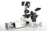 New Leica SD6000 Spinning Disk Confocal
Leica's new SD6000 Spinning Disk Confocal module is designed for ultra fast image acquisition with minimum sample exposure and is ideal for observing differentiation processes, intracellular transport, cell division, and dynamics of the cytoskeleton over long periods of time.
The new Leica SD6000 Spinning Disk Confocal utilizes software that guides users through experiments. Its streamlined user interface reduces the time spent hunting for functions in a menu bar and time spent training new users, which makes this imaging system very easy to use. In addition, the SD6000 does not require the use of a laser source. Rather, a ‘white light' source is used that is very simple and affordable to maintain.
The Leica SD6000 is designed around a fully automated, inverted, research microscope. As such, this system can utilize climate chambers and other live cell accessories to customize the solution to each laboratory's needs. In addition, the SD6000 can be retrofit to existing Leica AF6000 LX workstations and can be outfit new with Total Internal Reflection Fluorescence (TIRF) capability. All contrast techniques from Confocal to DIC to widefield fluorescence can be controlled automatically and integrated with the same experiment for complete flexibility.
In general, cellular events happen very quickly, in the order of milliseconds, and spinning disk technology provides a spectacular balance between speed and resolution to image these events. The Leica SD6000's rotating pinhole simultaneously scans all image points in the focal plane at a rate of 1000 times per second. The combination of this Multi-point Scanning and highly sensitive EMCCD fluorescence camera technology allows images to be recorded at the ultra high speed required for live cell research. By design, the SD6000 provides an instantly deblurred image on-screen as its pinholes quickly scan the field-of-view. This technique is much quicker than deconvolving widefield fluorescence images and allows real time imaging of biological processes.
The Leica SD6000 minimizes cell photo bleaching and photo toxicity, which enables biological processes to be observed over long periods of time. This is possible since fluorescence excitation via the spinning disk is a fraction of that used in widefield imaging. Therefore, cells can typically be studied for longer intervals in comparison. To ensure imaging sensitivity with lower excitation power, the SD6000 employs a highly sensitive, low-light, EMCCD fluorescence camera. Finally, fast filter wheels inside of the Confocal device further reduce specimen exposure times, which ensures sample protection for long-term studies.