A fluorescence microscope with structured lighting for quick super-resolution imaging across a large field of view has been created by researchers. The new microscope was developed to examine several living cells at once with very high resolution to study the impact of various medications and drug combinations on the body.
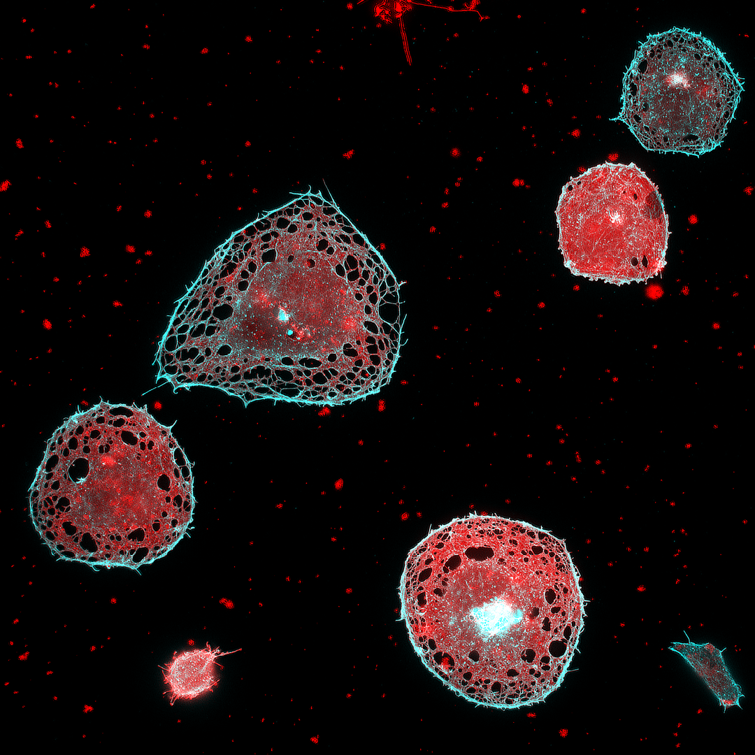 Thanks to the microscope’s large field of view, it is possible to acquire super-resolution images of multiple cells at once. Image Credit: Henning Ortkrass, Bielefeld University
Polypharmacy—the effect of the many combinations of drugs typically prescribed to the chronically sick or elderly—can lead to dangerous interactions and is becoming a major issue. We developed this microscope as part of the EIC Pathfinder OpenProject DeLIVERy, which aims to develop a platform that can investigate polypharmacy in individual patients.
Henning Ortkrass, Professor, Faculty of Physics, Bielefeld University
The researchers published their new microscope in the Optica Publishing Group’s journal Optics Express, which combines optical fiber transmission of excitation light to offer extremely high-quality images over a very broad field of view with multicolor and high-speed capabilities.
They demonstrate that the device can image liver cells with a field of view of up to 150×150 μm² and imaging speeds of up to 44 Hz while retaining a spatiotemporal resolution of less than 100 nm.
Ortkrass added, “With this new microscope, individual drug combinations can be tested on isolated cells and then imaged with super-resolution to observe dynamics of cell membrane features or organelles. The large field of view can provide statistical information about the cell response, which could be used to improve personalized healthcare. Thanks to the system’s potentially small size, it might also be useful for clinical applications where high resolution is important.”
High Resolution Across a Large Field of View
The new microscope is based on super-resolved structured illumination microscopy (SR-SIM), which employs a structured pattern of light to stimulate fluorescence in a sample and attain spatial resolution beyond the light diffraction limit. Since it employs low-power stimulation that does not damage the sample while delivering very detailed images, SR-SIM is particularly well suited for live cell imaging.
The new microscope reconstructs super-resolved images from a series of raw images to attain high resolution across a wide field of view. These raw images are obtained by illuminating the sample with a sinusoidal striped pattern that is shifted and rotated to get additional information using a set of six optical fibers. This results in a two-fold boost in resolution while still attaining quick imaging and being compatible with live-cell imaging.
“The fiber selection and phase shift are performed using a newly designed fiber switch based on galvanometric mirrors and MEMS-mirrors. We also custom-designed a hexagonal holder that collimates and refocuses the beams of the six fibers into the microscope to illuminate a large FOV and allow precise adjustment of all beams. This allows the setup to be used for total internal reflection fluorescence excitation (TIRF)-SIM, which is used to restrict fluorescence excitation and detection to a thin region of the sample,” further stated Ortkrass.
Imaging Liver Cells
Since the liver is the principal organ involved in drug metabolism, the researchers tested the system on samples of fixed multicolor-stained rat liver cells. The new microscope’s reconstructed images permitted the viewing of microscopic membrane structures smaller than the diffraction limit of light.
Ortkrass added, “This compact system uniquely combines a large field of view and fast pattern switching speed with multicolor, power-efficient excitation. In addition, the setup achieves very high image quality and can be tuned to perform either 2D-SIM or TIRF-SIM.”
The researchers want to use the microscope setup in the future to study the dynamics of cells treated with various medications in live cell investigations of liver cells. They also intend to enhance the image reconstruction process to achieve real-time reconstruction of the collected raw data.
Journal Reference:
Ortkrass, H., et al. (2023) High-speed TIRF and 2D super-resolution structured illumination microscopy with a large field of view based on fiber optic components. Optics Express. doi:10.1364/OE.495353
Source: https://www.optica.org/