In an article published in the journal Micromachines, researchers used a hyperspectral imaging system for the colorimetric characterization of four in-vivo maxillary anterior teeth. Results were compared with similar studies carried out with other measuring systems to validate the proposed capturing protocol.
Study: Validation of a Hyperspectral Imaging System for Color Measurement of In-Vivo Dental Structures. Image Credit: Alex Mit/Shutterstock.com
How Color of Human Teeth is Defined
Human teeth are made up of pulp (soft tissue) coated by two hard tissues, i.e. enamel and dentin. The optical characteristics of each of these tissues are quite distinctive, and they are also very complicated. Due to its translucency, dentine mostly determines tooth color, with enamel having a small impact. However, the ultimate shade of a tooth is partly influenced by the thickness of the enamel and dentine layers, which affects the amount of light that passes through them.
As the layer thickness changes depending on the different tooth sections rather than being constant across the whole tooth structure, it results in a color gradient along the various axes of the natural dental structures.
With standard instruments used in clinical settings, which often determine color by single spot measurements or broad, integrated measuring regions, these color changes inside tooth structures are exceedingly challenging to assess.
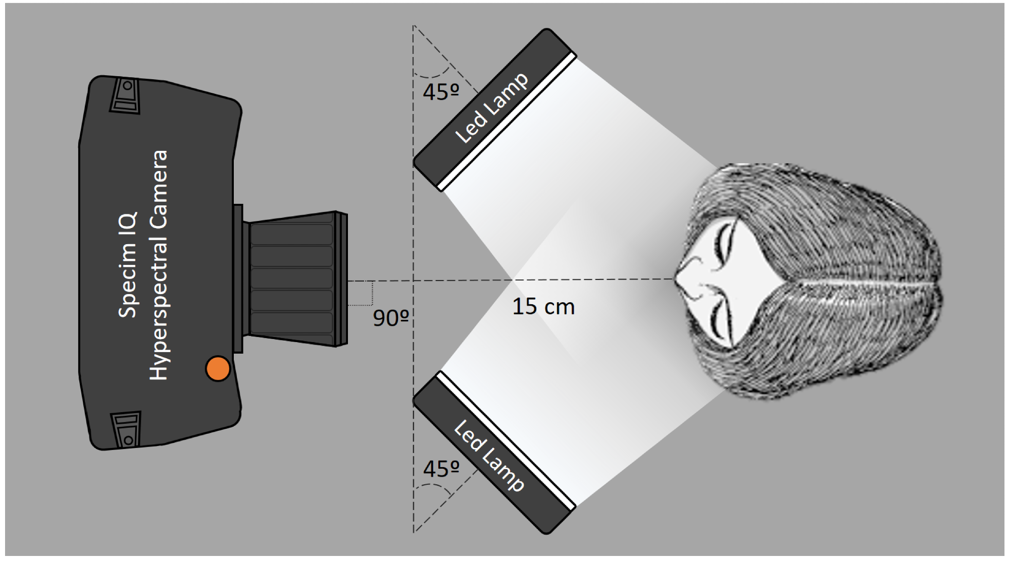
Figure 1. Schematic representation of the measurement set-up used to measure the hyperspectral reflectance images of the four maxillary incisors.
Tools for Color Measurements in Dentistry
For objective color measurements in dentistry, colorimeters, spectrophotometers, and digital imaging systems are now the most used tools. Each of these tools has benefits and drawbacks.
Colorimeters
Despite strong measurement reproducibility, colorimeters are susceptible to systematic errors brought on by edge-loss effects from the sample's surface.
Spectrophotometers
On the other hand, due to their capacity to detect the quantity of light reflected over the visible spectrum range, spectrophotometers may provide more systematic and accurate readings than colorimeters or digital imaging techniques, even if they need calibration for accurate measurement.
Digital Imaging Systems
Finally, digital imaging systems are the simplest method for measuring color in dentistry. They are becoming more common in the dental industry owing to the opportunities they open up when used with other software and editing tools. However, they are not systematized, need calibration, and involve the human eye in some degree of subjective shade choosing.
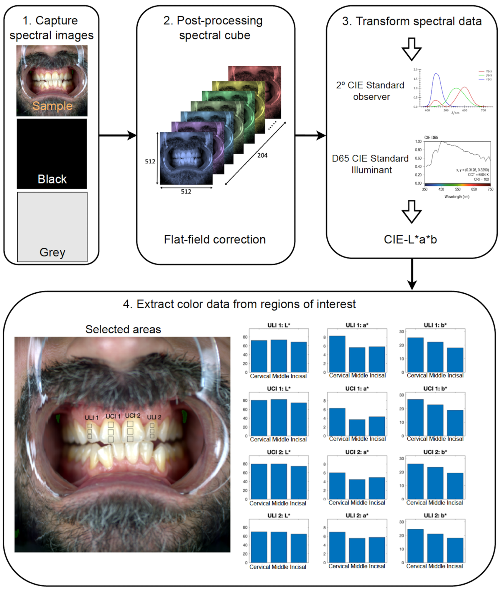
Figure 2. Workflow scheme of the spectral image capturing protocol, data processing and CIE L*, a* and b* values extraction corresponding to the cervical, middle and incisal thirds.
Correlations between Adjacent Teeth and Tooth Segments
Using various color measurement tools, including spectrophotometers, digital photography, and colorimeters, many researchers have sought to discover the color relationship between three tooth segments, i.e. the cervical, middle, and incisal. These researchers concluded that there are considerable variances between various teeth and sections of the same tooth and that to maximize the effectiveness of the restorations, unique shade selection for each tooth and different areas of a tooth should be taken into consideration.
Therefore by analyzing the correlations between contralateral and adjacent teeth as well as various areas of the same tooth, this study aimed to validate the use of a hyperspectral imaging device for in-vivo color measurement of the four maxillary anterior teeth. The results were then compared with those found in the current literature.
How the Study was Conducted
Choosing Participants
To participate in this study, 30 participants (50 percent males, 50 percent women, ranging in age from 21 to 67 years; average age, 29 years) were chosen. All participants were examined to determine the viability of the target teeth before beginning hyperspectral image acquisition.
Experimental Equipment and Procedure
A hyperspectral camera, two LED lights, and a chin rest made up the experimental apparatus. The camera was positioned 15 cm from the target tooth to maximize the size of the region being caught by the camera. In the end, the LED lamps were arranged so that it would be possible to achieve a CIE 45/0 illuminating/measuring geometry.
The line integration time was set at 40 milliseconds for each capture, corresponding to approximately 8 s of picture-taking time. The CIE 2 Standard Observer and the CIE D65 Standard Illuminant were used to translate the spectral reflectance data into CIELAB values. Each tooth was divided into the cervical third, middle third, and incisal third regions of interest to study the chromatic changes along the dental structures. The mean CIE L*, a*, and b* values were taken from these three regions.
The CIELAB (ΔE*ab) and CIEDE2000 (ΔE00) total color difference formulae were used to calculate the color differences between target teeth and tooth regions. In addition, the matching 50:50% perceptibility (PT) and acceptability (AT) criteria for the color difference in dentistry were used to assess (ΔE*ab) and (ΔE00) values.
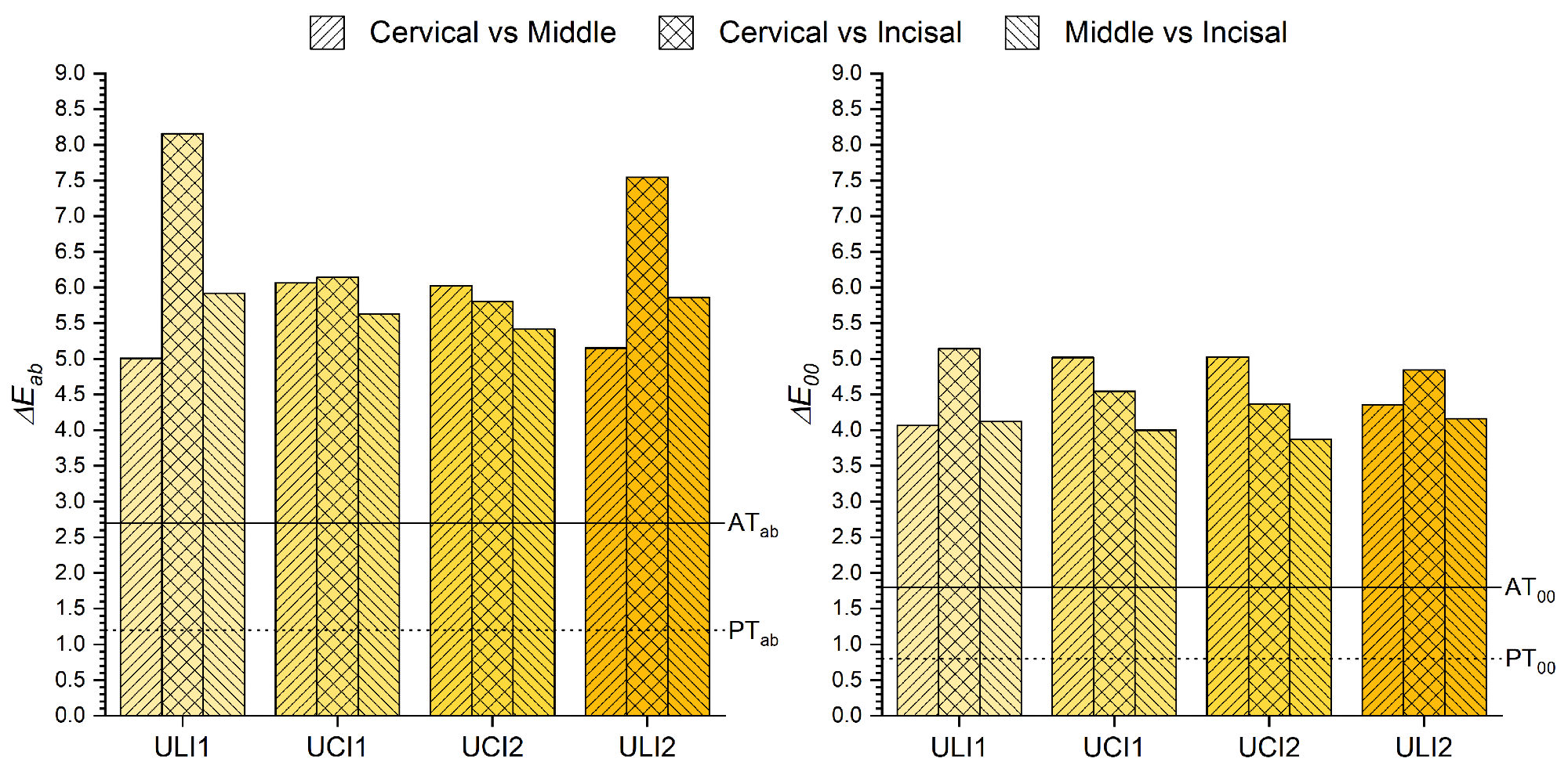
Figure 3. ΔE∗ab and ΔE00 color differences between the cervical, middle and incisal thirds of the same tooth.
Significant Findings of the Study
In every instance, the mean color differences between upper central (UCI) and lateral incisors (ULI) surpassed AT (ΔE00 = 5.71–5.74; ΔE*ab = 7.39–7.42). In addition, there were noticeable chromatic differences between the cervical (C), middle (M), and incisal (I) sections of the same tooth. (ΔE00 = 3.87–4.16 and ΔE*ab = 5.42–5.92; ΔE00 = 4.37–5.15 and ΔE*ab = 5.80–8.16; and ΔE00 = 4.07–5.03 and ΔE*ab = 5.01–6.07 between M and I, C and I and, C and M respectively).
Using a hyperspectral camera has shown to be a reliable and effective method for determining the spectral reflectance of in-vivo natural teeth within the constraints of this study.
Reference
Maria Tejada-Casado, Razvan Ghinea, Miguel Ángel Martínez-Domingo, María M. Pérez, Juan C. Cardona, Javier Ruiz-López and Luis Javier Herrera (2022) Validation of a Hyperspectral Imaging System for Color Measurement of In-Vivo Dental Structures. Micromachines. https://www.mdpi.com/2072-666X/13/11/1929/htm
Disclaimer: The views expressed here are those of the author expressed in their private capacity and do not necessarily represent the views of AZoM.com Limited T/A AZoNetwork the owner and operator of this website. This disclaimer forms part of the Terms and conditions of use of this website.