Sep 9 2022Reviewed by Mila Perera
How can a Ph.D. candidate with a fluorescence microscope and a live bacterial sample use these resources to get precise observations of bacterial division from the sample?
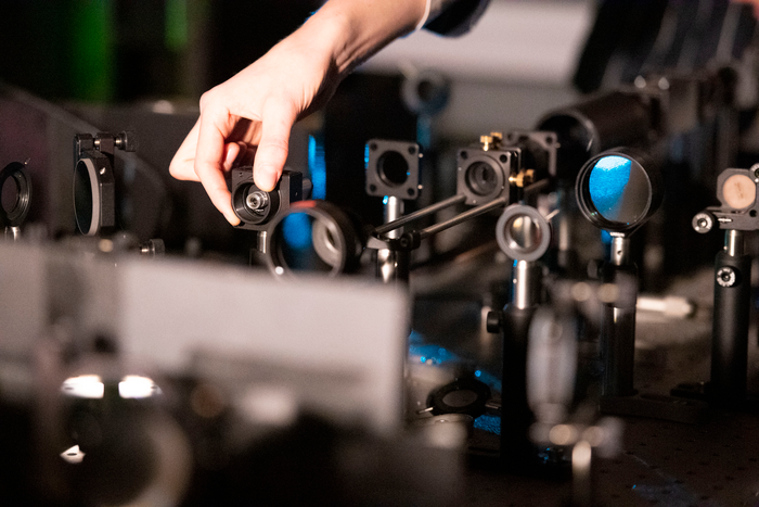 Suliana Manley’s fluorescent microscope at EPFL. Image Credit: Hillary Sanctuary/EPFL.
When bacterial division finally begins, a researcher can be tempted to skip meals and sleep (one microbe can divide for hours) to spend all of their time taking pictures under the microscope since manual detection and acquisition control is common in many fields.
An alternative is to program the microscope to take pictures as frequently and randomly as possible, but too much light speeds up the sample’s loss of fluorescence and risks the early demise of live samples. Furthermore, since only a small number of images would show bacteria in division, there is a possibility that numerous irrelevant photographs will be produced using this method.
Another approach is to utilize artificial intelligence to identify bacterial division precursors and use these to automatically update the control software of the microscope to capture images of the process.
With the use of artificial neural networks, EPFL biophysicists have discovered a method to automate microscope control for imaging biological activities in great detail while minimizing stress on the sample. Both bacterial cell division and mitochondrial division can be accomplished using their method. The study was published in Nature Methods journal.
An intelligent microscope is kind of like a self-driving car. It needs to process certain types of information, subtle patterns that it then responds to by changing its behavior. By using a neural network, we can detect much more subtle events and use them to drive changes in acquisition speed.
Suliana Manley, Principal Investigator, Laboratory of Experimental Biophysics, EPFL
Initially, Manley and her team discovered how to identify mitochondrial division, which was more challenging than for bacteria like C. crescentus. As mitochondrial division happens infrequently, it can happen at any time and almost anywhere in the mitochondrial network.
However, the researchers found the solution by training the neural network to look out for mitochondrial constrictions, a change in the shape of mitochondria that leads to division, and observations of a protein known to be plentiful in regions of division.
When both constrictions and protein levels are high, the microscope uses high-speed imaging to capture various detailed images of division events. When constriction and protein levels are low, the microscope switches to low-speed imaging to avoid overexposing the sample.
The researchers demonstrated that they could view the material for a more extended period of time with this intelligent fluorescent microscope than with conventional quick imaging. Even though the sample was under more stress than with slow imaging as is customary, they were able to gather more insightful information.
The potential of intelligent microscopy includes measuring what standard acquisitions would miss. We capture more events, measure smaller constrictions, and can follow each division in greater detail.
Suliana Manley, Principal Investigator, Laboratory of Experimental Biophysics, EPFL
The scientists are making the control framework available as an open source plug-in for the free microscope software Micro-Manager, allowing other researchers to add artificial intelligence to their own microscopes.
Journal Reference:
Mahecic, D., et al. (2022) Event-driven acquisition for content-enriched microscopy. Nature Methods. doi.org/10.1038/s41592-022-01589-x.
Source: https://www.epfl.ch/en/