Endoscopes are a common component of health checkups. The endoscope is a long, flexible tube that houses a camera and a light source for imaging purposes. It is especially helpful for getting pictures of the human body’s interior. For instance, endoscopies of the stomach and colon are frequently utilized to identify and diagnose disorders, including cancer and ulcers.
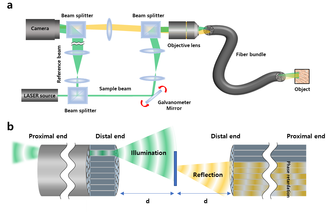 Experimental setup of ultrathin holographic endomicroscope. (a) The output beam from a laser is divided into sample and reference beams. The sample beam is delivered to the sample through the fiber bundle. The backscattering signal from the sample, indicated as yellow for clarity although its wavelength is identical to the incident wave, is captured by the fiber bundle and delivered to the camera. The reference beam generates an interferogram together with the signal beam at the camera. (b) Image formation principle. The angular spectrum of the sample is obtained under Fresnel conditions by separating the distance between the object and the optical fiber. Image Credit: Institute for Basic Science
Creating an endoscope often involves using an optical fiber, which enables information to be communicated using light, or connecting a camera sensor to the end of a probe. The thickness of the probe rises when an endoscope has a camera sensor, making the procedure more invasive.
An endoscope that uses an optical fiber bundle can be produced in a smaller form factor, minimizing invasiveness and causing the patients significantly less discomfort.
The drawback is that high-resolution imaging is challenging with a traditional fiber-bundle endoscope since the image's resolution is constrained by the size of the individual fiber cores.
The probe tip’s reflection also results in the loss of a significant amount of image data. Furthermore, due to the significant back-reflection noise produced by the tip of the thin probe during fiber endoscopy, it is frequently required to identify the target with fluorescence, particularly in biological substances with low reflectivity.
A high-resolution holographic endoscope system has recently been created by a research team under the direction of Wonshik Choi, Associate Director of the Center for Molecular Spectroscopy and Dynamics (CMSD) at the Institute for Basic Science (IBS).
Without anchoring a lens or any other equipment to the distal end of the fiber bundle, the researchers could get around the previous fiber optic endoscopy restriction and reconstruct high-resolution images.
By measuring the holographic representations of the light waves that are reflected from the object and gathered by the fiber bundle, this task was accomplished. Before measuring the holographic pictures that were reflected from the object at a specific distance from the optical fiber, the researchers lit the object by focusing light onto a single core of a fiber bundle.
By adjusting the phase retardation caused by each fiber core during the analysis of the holographic images, it was possible to rebuild the object image with microscopic detail.
To remove fiber-induced phase retardations in both the illumination and detection paths and rebuild an object image with microscopic resolution, a special coherent image optimization technique was created.
The probe of the developed endoscope is 350 μm in diameter, which is smaller than the needle used for hypodermic injection since it does not connect any equipment to the end of the optical fiber.
Through the use of this method, scientists were able to get high-resolution images with a spatial resolution of 850 nm, which is much lower than the core size of the optical fiber bundle.
The researchers then experimented with the brand-new Fourier holographic endoscopy technique to visualize the structure of mice villi. Even in biological samples with extremely low reflectivity, such as rat villi, excellent contrast images could be acquired by successfully eliminating the probe’s back-reflection noise.
Furthermore, using post-processing techniques on the measured holographic data, it was possible to create multi-depth 3D images from a single data set with a depth resolution of 14 μm.
This new endoscope is anticipated to significantly advance the ability to examine the internal organs of human bodies with minimally invasive procedures that cause patients little to no discomfort.
Additionally, it will allow for the direct observation of spaces as tiny as microvessels and the tiniest airways in the lungs, which was not conceivable with earlier technologies. Even while it could be useful for industrial inspections of semiconductors and microprocessors, researchers indicated that the applications of their new endoscope went far beyond the realm of healthcare.
On August 2nd, 2022, this research was published in Nature Communications.
Journal Reference:
Choi, W., et al. (2022) Flexible-type ultrathin holographic endoscope for microscopic imaging of unstained biological tissues. Nature Communications. doi:10.1038/s41467-022-32114-5.
Source: https://www.ibs.re.kr/eng.do