Apr 22 2022Reviewed by Alex Smith
Scientists have formulated a unique measurement and imaging method that can resolve nanostructures smaller than the diffraction limit of light without necessitating any labels or dyes.
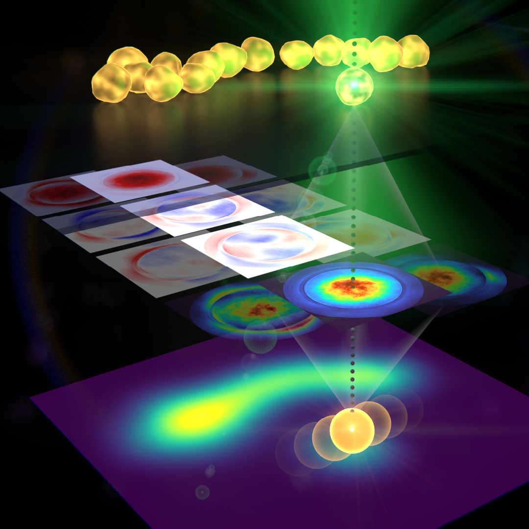 Researchers have developed a new measurement and imaging approach that can resolve nanostructures smaller than the diffraction limit of light. After light interacts with a sample, the new technique measures the light intensity as well as other parameters encoded in the light field. Image Credit: Jörg S. Eismann, University of Graz
The study signifies a noteworthy advance toward a new and robust microscopy technique that could soon be applied to observe the fine characteristics of complex samples, surpassing what is imaginable with traditional microscopes and methods.
Illustrated in Optica, Optica Publishing Group’s journal for highly influential research, the novel technique is a modified version of laser scanning microscopy, which uses a powerfully focused laser beam to irradiate a sample.
The scientists built on the method by computing not only the brightness, or concentration of the light after it interacts with a sample under investigation but also identifying other parameters encrypted in the light field.
Our approach could help extend the microscopy toolbox used to study nanostructures in a variety of samples. In comparison to super-resolution techniques based on a similar scanning approach, our method is fully non-invasive, meaning it doesn’t require any fluorescent molecules to be injected into a specimen before imaging.
Peter Banzer, Study Lead, University of Graz, Austria
The scientists demonstrate that they can compute the size and position of gold nanoparticles with an accuracy of several nanometers, even if numerous particles were touching.
“Our novel approach to laser-scanning microscopy could close the gap between conventional microscopes with limited resolution and super-resolution techniques that require modification of the specimen under study,” said Banzer.
Capturing More from Light
In laser-scanning microscopy, a beam of light is scanned across the sample, and the transmitted, scattered or reflected light emanating from the sample is measured. Although most microscopy approaches measure the brightness or intensity of light emanating from the sample, a great amount of data is also stored in other features of the light, for example, its polarization, phase and scattering angle.
To capture this extra data, the scientists inspected the spatial resolution of the intensity and polarization data.
The phase and polarization of light, together with its intensity, vary spatially in a way that incorporates fine details about the sample with which it interacts — much like the shadow of an object tells us something about the shape of the object itself. However, much of this information is ignored if only the overall light power is measured after the interaction.
Peter Banzer, Study Lead, University of Graz, Austria
They exhibited the new method by using it to examine basic samples comprising metallic nanoparticles of various sizes. They did this by scanning the region of interest and then recording polarization and angle-resolved images of the diffused light. The measured data was assessed using an algorithm that forms a model of the particles that automatically adjusts to resemble the measured data as precisely as possible.
“Although the particles and their distances were much smaller than the resolution limit of many microscopes, our method was able to resolve them,” said Banzer. “In addition, and even more importantly, the algorithm was able to provide other parameters about the sample such as the precise size and position of the particles.”
The scientists are currently involved in adapting the technique so that it could be applied to more intricate samples. The approach’s functionality can also be extended by customizing the structure of the light that interacts with the sample and integrating artificial intelligence-based methods into the image processing steps.
Regarding the detection side, the researchers, along with other professionals, are presently building a special camera as part of a European project referred to as SuperPixels. This advanced detection device will have the capacity to resolve polarization and phase information as well as intensity.
Our study is another demonstration of the pivotal role that the structure of light can play in the field of optics and light-based technologies. Many intriguing applications and phenomena have been demonstrated already, but there is more to come.
Peter Banzer, Study Lead, University of Graz, Austria
Journal Reference:
Eismann, J.S. & Banzer, P. (2022) Sub-diffraction-limit Fourier-plane laser scanning microscopy. Optica. doi.org/10.1364/OPTICA.450712.
Source: https://www.optica.org/en-us/home/