Mar 17 2021
A new intravascular imaging approach recently developed by scientists could someday be used for detecting coronary plaques that may trigger a heart attack.
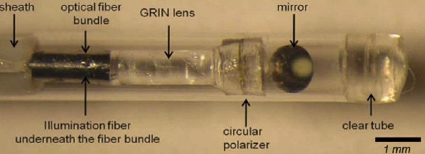 A new technique known as intravascular laser speckle imaging could one day be used to detect coronary plaques that are likely to lead to a heart attack. The researchers developed a small diameter intravascular catheter that incorporates a small-diameter fiber bundle, polarizer, and GRIN lens to image the reflected speckle patterns onto a CMOS sensor. Image Credit: Seemantini Nadkarni, Wellman Center for Photomedicine.
Generally, heart attacks occur when an unstable plaque bursts open and subsequently inhibits a crucial artery that transports oxygen and blood to the heart.
If unstable coronary plaques could be detected before they rupture, pharmacological or other treatments could be initiated early to prevent heart attacks and save lives. Our new imaging technique represents a major step toward achieving this.
Seemantini Nadkarni, Research Team Leader, Wellman Center for Photomedicine, Massachusetts General Hospital
In Biomedical Optics Express, the journal of The Optical Society (OSA), the team has recently reported a preclinical demonstration of their latest intravascular laser speckle imaging (ILSI) method in a live animal model.
For the first time, the researchers demonstrated that the ILSI technique can detect the clear mechanical properties of plaques that have a greater chance to rupture under physiological conditions of breathing, blood flow and cardiac motion.
Reducing mortality from heart attacks in the general population requires a comprehensive screening strategy to identify at-risk patients and detect high-risk vulnerable plaques while they can be treated. By providing the unique capability to measure mechanical stability—a critical metric in detecting unstable plaques—ILSI is poised to provide a new approach for coronary assessment.
Seemantini Nadkarni, Research Team Leader, Wellman Center for Photomedicine, Massachusetts General Hospital
Capturing Mechanical Stability of Plaques
While intravascular technologies have been designed to assess the microstructural characteristics of unstable plaques, new research works have demonstrated that mechanical traits, apart from compositional and microstructural features, have an impact on the rupture of plaques.
Measurement of the plaque mechanical properties is crucial in identifying unstable plaques with a propensity for rupture and subsequent heart attack. ILSI provides the unique capability to quantify an index of mechanical properties of coronary plaques, thus providing a direct assessment of mechanical stability.
Seemantini Nadkarni, Research Team Leader, Wellman Center for Photomedicine, Massachusetts General Hospital
To predict the mechanical properties, the ILSI technique makes use of laser speckle patterns that form when laser light is dispersed from tissues. When these speckles are visualized through a high speed camera, they vary in time because of the viscoelastic characteristics of the plaque.
This approach enables the team to quantify and differentiate the mechanical characteristics of unstable plaques, which have an abundant amount of lipids.
“For this new study, we developed a small diameter intravascular catheter that incorporates an optical fiber that delivers light to the coronary artery wall. We also used a small-diameter fiber bundle, polarizer, and GRIN lens to image the reflected speckle patterns onto a CMOS sensor,” added Nadkarni.
For preclinical testing, the investigators assessed the potential of their new ILSI device to identify the presence of unstable plaques in a human coronary to swine xenograft model system. A model system like this employs human coronary arteries that are stitched to the beating heart of a live, anesthetized pig.
The researchers evaluated the mechanical features of the plaque within the arteries by estimating the time constant, or rate, of variations in the intensity of the speckle patterns and subsequently compared their outcomes with histopathological results.
Nadkarni added, “The time constants in unstable plaques were significantly and distinctly lower than other stable plaques in the coronary wall. These results demonstrated the exquisite diagnostic sensitivity and specificity of ILSI for detecting human lipid pool plaques that were most likely to rupture under physiological conditions.”
According to the investigators, the novel method could be effortlessly incorporated into other intracoronary technologies, like intravascular ultrasound or optical coherence tomography, to merge the mechanical results from the ILSI technique with the morphological data and thus improve the assessment of the stability of plaques.
The team has planned to continue to investigate the ability of their new ILSI instrument for rapid evaluation of the coronary vasculature in live animals.
After completing these preclinical research works, they will evaluate the safety of the catheter for use in human beings and will subsequently initiate the process of obtaining regulatory approval for clinical applications.
Journal Reference:
Hajjarian, Z., et al. (2021) In-vivo mechanical characterization of coronary atherosclerotic plaques in living swine using intravascular laser speckle imaging. Biomedical Optics Express. doi.org/10.1364/BOE.418939.
Source: https://www.osa.org/