Dec 19 2019
Sechenov University researchers, in association with their collaborators from Australia, have suggested a new, cheaper, and faster way to determine the quality of meat. The method involves exposing a small amount of sample to UV light and quantifying the emission spectrum.
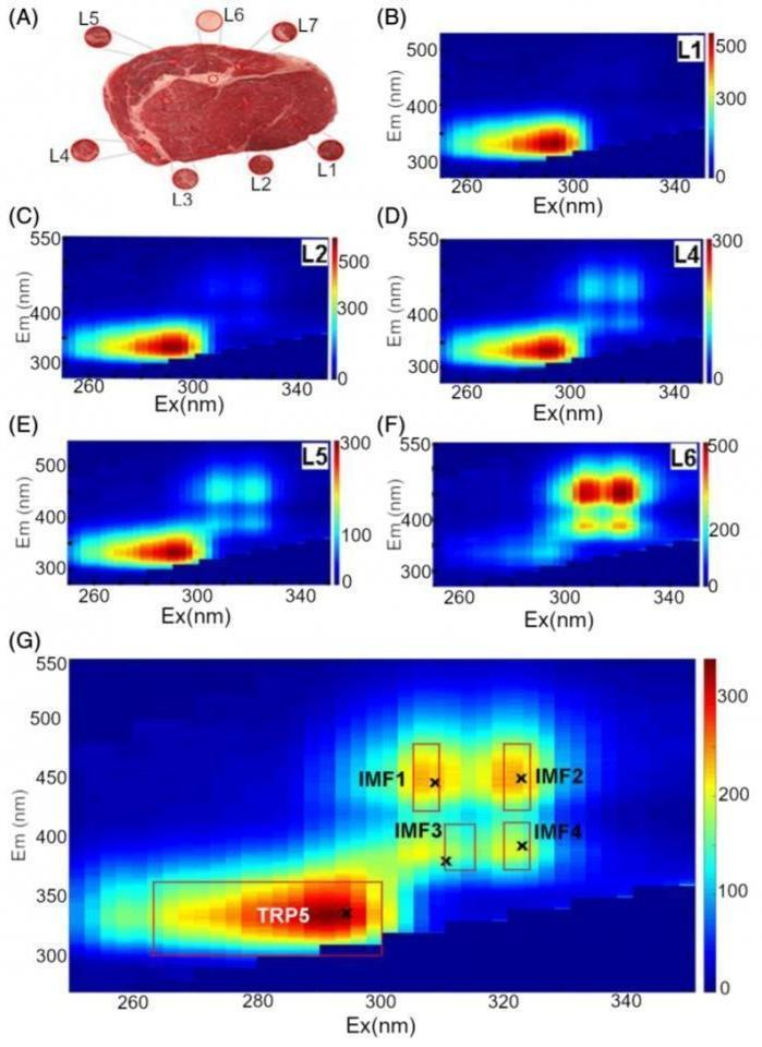 Matrices showing the intensity of fluorescence with various frequencies of excitation and emission. On the last one areas congruent to spectra of intramuscular fat (IMF) and amino acid tryptophan (TRP) are highlighted. Image Credit: Islam K et al. Autofluorescence excitation-emission matrices as a quantitative tool for the assessment of meat quality. J. Biophotonics. 2019; e201900237. © 2019 Wiley-VCH Verlag GmbH & Co. KGaA.
It has been demonstrated that the technique can accurately classify meat into usual quality categories. The method and the outcomes of the study have been described in the Journal of Biophotonics.
Specialists traditionally assess the quality of beef by focusing on its color, carcass weight, pattern of the fibers (marbling), etc. However, these measurements take plenty of time and largely depend on the experts’ subjective opinion.
An alternative method could be fluorescence spectroscopy. This method helps in identifying and quantifying the concentration of several compounds that can emit light of a particular frequency range.
Such substances contain several organic molecules that are present in meat. Previous studies have elucidated the fluorescence spectrum of a few ingredients of meat (numerous cell types of muscle, connective tissue, and adipose (fat)). This data was used by a number of scientific groups to evaluate the product’s specific characteristics, for example, the percentage of fatty acids or connective tissue.
The authors of the article published in the Journal of Biophotonics mapped the fluorescence spectrum of meat with its quality defined by three categories, such as MSA3, MSA4, or MSA5. The outcomes were further verified through histological (tissue and cell) analysis of the samples and quantifying both fat and water concentrations present in the samples.
The researchers utilized five pieces of meat in their study, for each of the three classes: Among the qualified meat types, the MSA3 class marks the lowest quality slices, while the MSA5 marks the highest quality ones. Six samples, each measuring approximately 8 mm in diameter, were cut from different areas of the meat steaks, where the relative content of muscle and fat tissues differed.
The scientists subsequently exposed the samples to light with a wavelength of 250–350 nm (middle and near-ultraviolet) and quantified the fluorescence spectrum ranging from 285 to 635 nm (from the center ultraviolet to the border between infrared and visible light). The emission intensity was fixed on the matrix “frequency of excitation - frequency of emission.”
The findings demonstrated that it is possible to perceive the fluorescence spectra of the samples with numerous ratios of adipose and muscle tissues. Spots present on the matrices of the adipose tissue samples can be distinguished and these spots correspond with the fluorescence spectrum of fat-soluble vitamins (A, D, K1, K2, and K3) as well as vitamin B and its components.
At the same time, the spectrum of the muscle tissue samples coincides with the spectrum of amino acid tryptophan it contains.
The features chosen by the authors allowed them to define the category of all pieces of meat. For instance, the highest quality meat—that is, MSA5—has the most intensive fluorescence and can be differentiated from samples of lower quality by the variation in brightness of different ranges.
The resultant data is also in agreement with the assumption that the existence of adipose and connective tissue is responsible for making the meat tender, while fat is responsible for its marbling.
This work shows the new opportunities to evaluate the quality of meat objectively by LED illumination and registration of the tissue optical response. It’s interesting to note that this technology, having being originally developed for the meat industry, can be further translated into medicine and biomedical research.
Dr Anna Guller, Study Co-Author and Senior Research Fellow, Sechenov University
She continued, “The principle on which this study was based, i.e. the detection of specific autofluorescence of various tissue components, allows evaluation of the structure and functional state of tissues without taking tissue fragments for biochemical of histological analysis.”
Therefore, our study can be considered as a possible step towards non-invasive and pain free diagnostics in medicine as well.
Dr Anna Guller, Study Co-Author and Senior Research Fellow, Sechenov University
Source: https://www.sechenov.ru/eng/