Jul 18 2019
Thanks to a new breakthrough, endoscopes may no longer have to be inserted into the body—for example, under the skin or down the throat—to reach the brain, stomach, or any other organs for inspection.
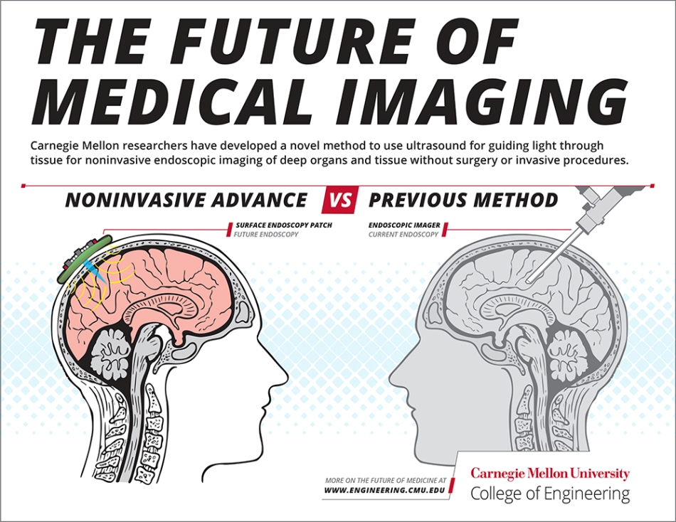 A noninvasive skin patch is one way the researchers’ ultrasound-based method could allow for easy imaging of the brain. (Image credit: Carnegie Mellon University College of Engineering)
A new method developed by Maysam Chamanzar, assistant professor of electrical and computer engineering, and Matteo Giuseppe Scopelliti, an ECE PhD student, utilizes ultrasound to noninvasively capture optical images via a turbid medium—for instance, biological tissue—to image the organs of the body. This novel technique can potentially remove the need for invasive visual exams utilizing endoscopic cameras.
Endoscopic imaging is an invasive procedure used for investigating and diagnosing symptoms of deep tissue disease. In this procedure, cameras are directly inserted into the body’s organs to examine symptoms. Cameras, or endoscopic imagers, provided on the end of catheter wires or tubes, are often implanted via a medical surgery or procedure to reach the deep tissues in the body. The new method developed by Chamanzar offers a fully noninvasive and non-surgical alternative.
The laboratory’s paper has been reported in Light: Science and Applications, a journal published by Springer Nature. It demonstrates that ultrasound can be used for producing a virtual “lens” inside the body, instead of implanting a physical lens. The use of ultrasonic wave patterns will allow the scientists to effectively “focus” light into the tissue, which enables them to capture images that can never be accessed before through noninvasive techniques.
Most of the light, specifically light in the optical spectrum’s visible range, is blocked by biological tissue. Hence, light cannot be used by present optical imaging techniques to access deep tissue from the surface.
However, noninvasive ultrasound is used by Chamanzar’s laboratory to induce greater transparency and thus allow more penetration of light via the turbid media, like biological tissue.
Being able to relay images from organs, such as the brain, without the need to insert physical optical components will provide an important alternative to implanting invasive endoscopes in the body. We used ultrasound waves to sculpt a virtual optical relay lens within a given target medium, which for example, can be biological tissue. Therefore, the tissue is turned into a lens that helps us capture and relay the images of deeper structures.
Maysam Chamanzar, Assistant Professor, Electrical and Computer Engineering, Carnegie Mellon University
“This method can revolutionize the field of biomedical imaging,” added Chamanzar.
Regardless of the type of medium they are flowing through, ultrasound waves can compress and rarefy. When compared to rarefied regions, light travels more slowly in compressed regions.
In this analysis, the researchers demonstrated that this effect of compression and rarefication can be used for molding a virtual lens in the target medium for optical imaging. It is possible to move this virtual lens around without disturbing the medium, and this is achieved by simply reconfiguring the ultrasound waves from the exterior. This makes it possible to image varied target regions in a fully non-invasive manner.
The published technique is a platform technology that can be used in a wide range of applications. In the coming days, the technique can be used in the form of a wearable surface patch or handheld device, based on the body’s organ being imaged.
Placing the patch or device on the skin would allow the clinician to effortlessly receive optical data from inside the tissue to produce images of what is inside without the various side effects and discomforts associated with endoscopy.
Endoscopic imaging under the skin or imaging of brain tissue is the closest present applications for this technology. However, this method can also be utilized in other parts of the body for imaging. This technique, in addition to biomedical applications, can be utilized in metrology, optical imaging in machine vision, and other industrial applications to facilitate steerable and non-destructive imaging of structures and objects at the micron scale.
The scientists demonstrated that the virtual “lens” properties can be adjusted by altering the parameters of the ultrasonic waves. This enables users to “focus” the captured images by using the technique at varied depths via the medium.
Although the LSA paper is targeting on the efficacy of the technique for closer-to-the-surface applications, the researchers are yet to identify the limit this ultrasonically-assisted optical imaging method can reach deep inside the body’s tissue.
What distinguishes our work from conventional acousto-optic methods is that we are using the target medium itself, which can be biological tissue, to affect light as it propagates through the medium. This in situ interaction provides opportunities to counterbalance the non-idealities that disturb the trajectory of light.
Maysam Chamanzar, Assistant Professor, Electrical and Computer Engineering, Carnegie Mellon University
The novel method could be used in several clinical applications, for example, monitoring brain activity, diagnosing skin disease, and diagnosis and photodynamic therapy for detecting and targeting malignant tumors.
Besides the direct applications on clinical medicine, the research will also have indirect clinical implications. Applying this acousto-optic technology to selectively activate different neural pathways and visualize mouse models of brain disorders in action would allow scientists to examine the mechanisms implicated in disease conditions like Parkinson’s. This can help in designing innovative clinical therapeutic interventions to treat such kinds of diseases in humans.
Turbid media have always been considered obstacles for optical imaging. But we have shown that such media can be converted to allies to help light reach the desired target. When we activate ultrasound with the proper pattern, the turbid medium becomes immediately transparent. It is exciting to think about the potential impact of this method on a wide range of fields, from biomedical applications to computer vision.
Matteo Giuseppe Scopelliti, PhD student, Electrical and Computer Engineering, Carnegie Mellon University
The scientists believe that within the next five years, this latest imaging technology could be used in clinical and biomedical contexts.
Maysam Chamanzar: Using Light to Enlighten Brain Function
(Video credit: Carnegie Mellon University College of Engineering)
Source: https://engineering.cmu.edu/