Feb 13 2019
Smartphones that are computationally powerful, accessible, and connected are not just for “selfies” anymore, but much more than that.
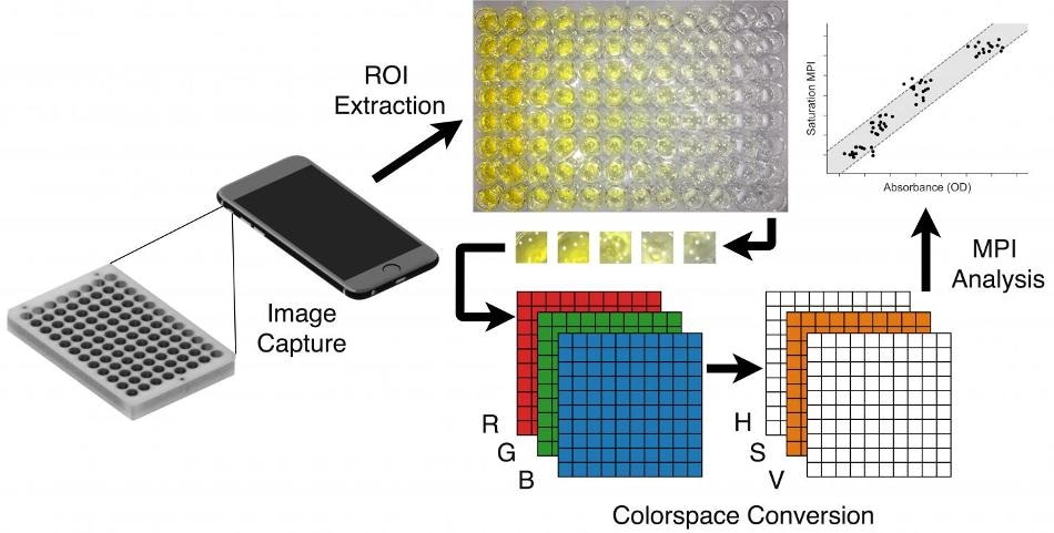 Images of a diagnostic assay are captured using a smartphone camera. Regions of interest are extracted and are converted to HSV (hue, saturation, value) space. After the conversion process, the standard pixel intensity analysis is applied to the saturation channel and the values are used to determine absorbance and concentration of the sample automatically. (Image credit: Florida Atlantic University)
Smartphones have evolved as robust evaluation tools that have the potential to diagnose medical disorders in point-of-care settings. Since smartphones enable even untrained users to obtain and relay data to medical professionals, they also offer a practical solution for health care in the emerging world.
Currently, smartphone camera technology provides a host of medical applications, for example, cytometric analysis and microscopy; however, in reality, cell phone image tests have some restrictions that considerably limit their application. To deal with these restrictions, external smartphone hardware is required to achieve quantitative outcomes—resulting in a design tradeoff between accuracy and accessibility.
At Florida Atlantic University’s College of Engineering and Computer Science, researchers have devised an innovative cell phone imaging algorithm that makes it possible to analyze assays, which are usually assessed through spectroscopy—a robust and highly advanced device employed in scientific research.
Using the analysis of over 10,000 images, the researchers were able to show that the saturation technique developed by them consistently surpassed the present-day algorithms under many different operating field conditions. The team’s findings have been reported in the journal Analyst of the Royal Society of Chemistry and represent a step ahead in advancing point-of-care diagnostics by enhancing the limit of detection, lowering the demand for required equipment, and boosting the accuracy of quantitative outcomes.
Smartphone cameras are optimized for image appearance rather than for quantitative image-based measurements, and they can't be bypassed or reversed easily. Furthermore, most lab-based biological and biochemical assays still lack a robust and repeatable cell phone analogue. We have been able to develop a cell phone-based image preprocessing method that produces a mean pixel intensity with smaller variances, lower limits-of-detection, and a higher dynamic range than existing methods.
Waseem Asghar, PhD, Study Lead Author and Assistant Professor, Department of Computer and Electrical Engineering and Computer Science, Florida Atlantic University.
For the analysis, Asghar, co-authors Chad Coarsey and Benjamin Coleman, and graduate students in the Asghar Laboratory in College of Engineering and Computer Science of FAU, used three smartphones—the iPhone 6 with a 12 megapixel (MP) camera, the Android Moto G with a 5 MP camera, and the Samsung Galaxy Edge 7 with a 12 MP camera—to capture the images.
Image capture was tested at numerous conditions, algorithm performance was measured, sensitivity to the distance, tilt, and motion of the camera was tested, and concentration-response and histogram properties were investigated. In addition, the researchers analyzed limit-of-detection and also ambient lighting levels, saturation properties, and relationship with red-green-blue, or RGB, color space. Images captured with a cell phone are natively preserved as arrays of RGB pixel intensities, often known as color channels.
When the researchers used several thousand images to compare saturation analysis with current RGB techniques, they observed that it empirically as well as analytically enhanced performance in the presence of multiplicative and additive ambient light noise. The team also demonstrated that saturation analysis can be understood as an improved version of prevailing RGB ratio tests. The researchers confirmed that conditions for the ideal image capture include a clean white background, a constant white light, the camera’s zero angular displacement, and minimal distance to the sample.
The trio—Coarsey, Asghar, and Coleman—even used the test on an enzyme-linked immunosorbent assay (ELISA), a plate-based assay method developed for identifying and measuring substances like hormones, antibodies¸ proteins, and peptides. The team found that saturation analysis for HIV allowed for an equipment-free assessment, and a limit-of-detection was also considerably lower compared to what is presently available with RGB techniques.
The methodology developed by FAU indicates an enhancement in practicality, repeatability, and rejection of image capture noise. Saturation analysis is also unaffected by most of the significant restricting factors for tests based on images, for example, shading, variations in ambient lighting, and fluctuating light levels. The investigators expect that the satisfactory characteristics of saturation analysis will encounter and allow for point-of-care tests based on cell phone images with lower limits-of-detection and less equipment overhead.
The research taking place in the Asghar Laboratory at Florida Atlantic University has important implications for diagnostic medicine and the delivery of health care in developed as well as developing countries. Professor Asghar and his team are driven to continue to develop cutting-edge technology that has the ability to remotely detect and diagnose diseases rapidly, accurately and inexpensively. This latest algorithm they have developed is one of the many advances they are making in this field.
Stella Batalama, PhD and Dean, College of Engineering and Computer Science, Florida Atlantic University.