Neuroimaging reveals the structure and function of the human brain. Techniques such as MRI, fMRI, CT, PET, EEG, and others have been fundamental in developing our knowledge of how the brain works and how it is altered in diseases that impact the brain, such as neurodegenerative disease (e.g. Alzheimer’s disease, Parkinson’s disease).
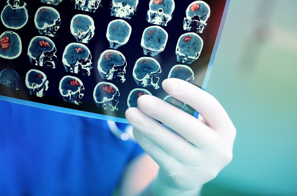
Image Credit: sfam_photo/Shutterstock.com
It has been vital in shedding light on how the brain works in mental health disorders (e.g., depression, anxiety, addiction, schizophrenia). As a result, neuroimaging has helped us to innovate novel ways to approach disease treatment, which has led to the more effective treatment of patients with a wide range of diseases.
Developments in optical technology are influencing the capabilities of neuroimaging platforms. The evolution of optics is benefiting the field of neuroimaging, pushing it further than ever before to help it achieve its full potential. Here, we discuss how this is happening in the specific instance of leveraging developments in multimode optical fibers. We reflect on how this may influence future results in neuroimaging and why this is important to health overall.
What are Multimode Optical Fibers?
Multimode fibers are optical fibers specifically designed to transport multiple light rays or modes of light simultaneously. The optical fiber core of these fibers carries each ray or mode at a fractionally different reflection angle, making it possible to transport many simultaneously.
It is generally accepted that multimode optical fibers are currently the most diffused tool that neuroscience laboratories have to interface with the brain’s deeper regions inaccessible to conventional imaging techniques such as EEG and fMRI.
While multimode optical fibers are often considered very limited systems, recent neuroimaging studies have revealed that advances in optics and photonics can be leveraged into neuroscience research, overcoming the limitations of traditional systems with multimode optical fibers.
How are Multimode Optical Fibers Being Used in Neuroimaging?
Multimode optical fibers are helping to enhance current neuroimaging techniques, allowing them to overcome current limitations so that we can gather more reliable, in-depth data on the workings of the human brain. With this data, we should be able to vastly expand our understanding of the human brain, and, therefore, develop novel approaches to treating neurological diseases.
A paper published in the journal APL Photonics in 2022 illustrates how multimode optical fibers are being leveraged to improve imaging techniques employed to visualize the brain’s subcellular structures that are notoriously difficult to study.
Miniaturized micro endoscopes are typically utilized to visualize the brain's deep structures. However, this method is limited in that it relies on gradient index (GRIN) microlenses that suffer from increasing aberrations and restricted fields of view as they are developed to become smaller and less invasive. This major limitation prevents them from visualizing the brain’s deep structures in detail necessary to reveal critical structural and functional information.
Researchers in the Netherlands overcame this limitation by utilizing multimode optical fibers within their endoscopes combined with advanced wavefront engineering techniques, allowing them access to deep tissue with minimal invasiveness due to high-resolution imaging. This research is the first time this concept was successfully demonstrated.
The team used auto-fluorescence human brain imaging via a single 50 μm-core multimode optical fiber probe. The team tested two approaches: an active wavefront shaping approach that acquired images by raster-scan imaging and a computational image recovery approach that used speckle-based compressive imaging technology.
The researchers reported that the approach using compressive imaging resulted in a significant decrease in image acquisition time for an area of interest up to three times larger than conventional techniques allow while maintaining high levels of spatial resolution.
Their results demonstrated lipofuscin accumulation, the age-related pigment implicated in Alzheimer’s disease. The visualization captured was 18 times faster than conventional methods.
Why is Neuroimaging Important and How Will it Evolve in the Future?
The multimode optical fiber method described in the research published in APL Photonics demonstrates a minimally invasive solution to imaging deep brain structures.
The proposed method offers a rapid, highly sensitive, and high-resolution imaging technique via apparatus that measures as thin as a single hair fiber.
The adaptation of this method will likely be vital in developing sensitive and effective ways of imaging delicate and difficult-to-visualize biological surroundings, such as deep brain tissue, internal organs, and other biomedical deep tissues.
Other neuroimaging and clinical research fields will likely benefit from this technique's development and the following advances. It is possible that, in the coming years, this method will accelerate developments in the field of neuroimaging, as it offers a way to capture data on hard-to-study areas.
As a result, we may see the emergence of novel approaches to treating neurodegenerative diseases, such as Alzheimer’s disease, and growing our understanding of the neural correlates of mental health illnesses.
References and Further Reading
De Vittorio, M. and Pisanello, F., 2021. Multimode Optical Fibers for Optical Neural Interfaces. Advances in Experimental Medicine and Biology, pp.565-583. https://pubmed.ncbi.nlm.nih.gov/33398843/
Lochocki, B., Verweg, M., Hoozemans, J., de Boer, J. and Amitonova, L., 2022. Epi-fluorescence imaging of the human brain through a multimode fiber. APL Photonics, 7(7), p.071301. https://aip.scitation.org/doi/10.1063/5.0080672
Turtaev, S., Leite, I., Altwegg-Boussac, T., Pakan, J., Rochefort, N. and Čižmár, T., 2018. High-fidelity multimode fibre-based endoscopy for deep brain in vivo imaging. Light: Science & Applications, 7(1). https://www.nature.com/articles/s41377-018-0094-x
Disclaimer: The views expressed here are those of the author expressed in their private capacity and do not necessarily represent the views of AZoM.com Limited T/A AZoNetwork the owner and operator of this website. This disclaimer forms part of the Terms and conditions of use of this website.