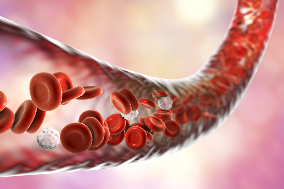
Image Credit: Kateryna Kon/Shutterstock.com
Three scientists based at POSTECH in Korea have enhanced the photoacoustic microscopy method, creating new 'super-resolution' photoacoustic microscopy that they have proven to be 500 times fast than the conventional method, with a resolution that is 2.5 times better.
The system has the capabilities of imaging blood vessels within the body in real-time with such precision that it could be developed for use in the diagnosis of stroke and cardiovascular disease, and is likely to find applications in vascular disease and hemodynamics.
The Benefits of Enhancing 'Gold Standard' Photoacoustic System
Until now, photoacoustic microscopy (PAM) has been considered the ‘gold standard’ when it comes to generating anatomical, molecular, and functional information about the human body. First used in the 1970s, the technique is a form of non-destructive testing that acoustically detects optical contrast via the photoacoustic effect.
Since its conception, PAM has significantly impacted a wide range of disciplines, including biomedicine, oncology, ophthalmology, neuroimaging, cardiology, even engineering. This is because of its advantages over other imaging techniques, such as it providing high-resolution images without the need for invasive tracers, and can concurrently image anatomical, functional, flow dynamic, metabolic, and molecular contrasts in vivo.
The Korean scientists saw an opportunity to enhance the PAM system. As it is relied upon by so many scientific fields, a marked improvement in the system would have a significant impact on the wide range of applications it has been developed for use in since the 1970s.
Chulhong Kim, who is the Professor of Creative IT Engineering at POSTECH, along with research professor Jinyoung Kim, and Jongbeom Kim, a Ph.D. student, described how they enhanced the PAM system in a paper published this month in the journal Nature, Light: Science and Applications.
Biological Tissue Research Using PAM
PAM sends optical energy as a laser beam towards biological tissues, which are then converted into heat energy when the tissues absorb the light, which initiates signature vibrations. PAM recreates imagery of what is happening inside the tissue, which could visualize cells, blood vessels, and other tissues.
Conventional PAM systems are limited in their field of view because they use galvanometer scanners that can only measure the optical beam and not the photoacoustic waves. They also have a temporal limitation in generating visualizations.
The POSTECH researchers developed an enhanced version of the conventional PAM system which resulted in improved performances. They created an agent-free localization approach that was based on a stable and commercial galvanometer scanner that had the added feature of a custom-made scanning mirror.
The research demonstrated that the new system significantly enhanced the temporal resolution while simultaneously preserving a high signal-to-noise ratio. The system they built can scan photoacoustic waves and optical beams concurrently due to the additional hardware of the custom-made scanning mirror.
Results also showed that the new system visualized even the very small blood cells using red blood cells alone without the addition of a contrast absorber. Also, the team showed that the system is 500 times faster, and has a super-resolution that is 2.5 times better than the conventional system.
A System that Images Even the Smallest Blood Vessels
A newly developed form of photoacoustic microscopy that utilizes a commercial galvanometer scanner alongside a custom-made scanning mirror was established by the team of scientists from POSTECH. Early investigations have demonstrated that it can successfully recognize blocked or burst blood vessels.
What was established in this study will be significant to various fields of science. Most importantly, it offers a potential new tool for the diagnosis and treatment of stroke and cardiovascular disease. It also has the potential to be used to vascular disease, to quickly and efficiently detect problems occurring within blood vessels. Finally, it has also been shown to be successful in the monitoring of hemodynamics in the microvessels.
It is expected that the system will be developed for use in a range of medical applications, as well as in preclinical and clinical studies.
References and Further Reading
- Kim, J., Kim, J., Jeon, S., BAIK, J., Cho, S. and Kim, C. (2019). Super-resolution localization photoacoustic microscopy using intrinsic red blood cells as contrast absorbers. Light: Science & Applications, 8(1). https://www.nature.com/articles/s41377-019-0220-4
- Yao, J. and Wang, L. (2013). Photoacoustic microscopy. Laser & Photonics Reviews, 7(5), pp.758-778. https://www.ncbi.nlm.nih.gov/pmc/articles/PMC3887369/
- Zhang, H. and Razansky, D. (2016). Special issue introduction: Photoacoustic microscopy. Photoacoustics, 4(3), pp.81-82. https://www.ncbi.nlm.nih.gov/pmc/articles/PMC5063352/
Disclaimer: The views expressed here are those of the author expressed in their private capacity and do not necessarily represent the views of AZoM.com Limited T/A AZoNetwork the owner and operator of this website. This disclaimer forms part of the Terms and conditions of use of this website.