Aug 6 2020
A new high-resolution X-ray imaging method recently demonstrated by scientists is capable of capturing the movement of rapidly changing dynamics and quickly moving objects.
 Researchers developed a high-resolution x-ray imaging technique based on ghost imaging that can capture the motion of rapidly moving objects. They used it to create a movie of a blade rotating at 100,000 frames per second. Image Credit: Sharon Shwartz, Bar-Ilan University.
This latest technique could potentially be used for imaging moving mechanical components in a non-destructive way and for capturing biological processes that were not formerly available with medical x-ray imaging.
The technique we demonstrated can be used with any x-ray source, plus it is low cost, simple and robust. Thus, it opens up the possibility of using x-rays to measure fast dynamics outside the lab.
Sharon Shwartz, Study Team Leader, Bar-Ilan University
The team has described the novel X-ray imaging technique in Optics Express—a journal from The Optical Society (OSA). In this method, a non-traditional imaging technique called ghost imaging is used to obtain rapid imaging speeds with excellent spatial resolution.
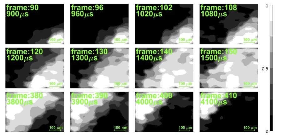
The researchers demonstrated the new method by producing an X-ray film of a blade that spins at 100,000 frames per second.
Medical imaging systems based on this technique could offer a new diagnostic tool for physicians. Our approach could, for example, be used to acquire high-resolution movies of the heart while greatly reducing the radiation dose for patients.
Sharon Shwartz, Study Team Leader, Bar-Ilan University
Seeing Through Surfaces
X-rays have a special capability to enter surfaces that are generally opaque to visible wavelengths, and hence, they are handy for imaging applications. Conventional X-ray imaging usually employs a pixelated camera with every pixel quantifying the level of intensity of the X-ray beam at a particular place.
More pixels are needed to capture higher-resolution X-ray images but this approach generates large amounts of information that take plenty of time to transfer. This leads to a trade-off between spatial resolution and imaging speed that renders it impossible to record high-speed events with excellent resolution.
While this trade-off can be resolved by highly specialized methods that involve extremely robust X-rays, such X-ray sources can be found only at huge synchrotrons that are installed at a few facilities worldwide.
In the latest study, the team used ghost imaging because this method employs single-pixel detectors that can enhance the speed of imaging. Ghost imaging typically functions by correlating a pair of beams—that is, X-ray beams, in this case. These beams do not separately carry any meaningful data relating to the object.
While one beam encodes a haphazard pattern that serves as a reference point and never probes the sample directly, the other one travels via the sample. In ghost imaging, only a minimal amount of X-ray power makes contact with the object being imaged, and hence, this method can also help decrease exposure to X-rays when employed for medical imaging.
Although single-pixel detectors can be much faster than pixelated detectors, they do not provide the spatial resolution necessary for image reconstruction. We used ghost imaging to overcome this problem and showed that we can image fast dynamics with spatial resolution comparable to or even better than the state-of-the-art x-ray pixelated detectors.
Sharon Shwartz, Study Team Leader, Bar-Ilan University
A Simple Solution
The researchers produced the reference beam required for ghost imaging by using regular sandpaper. The sandpaper was mounted on motorized stages to produce a haphazard pattern that was captured with a slow frame rate, high-resolution pixelated X-ray camera.
When the stage was shifted to different positions, the beam of X-rays bombarded a different section of the sandpaper, producing intensity fluctuations or haphazard X-ray transmissions. The pixelated camera was subsequently removed from the X-ray beam and a single-pixel detector as well as the object to be imaged was inserted.
The researchers then shifted the motorized stages to illuminate the object with the patterns of intensity fluctuations that were introduced at the numerous positions of the sandpaper and subsequently quantified the overall intensity after the X-ray beam bombarded the object with the single-pixel detector.
To apply this method to image a rapidly moving blade, the team synchronized the measurements with the movement of the blade. An ultimate image could be subsequently rebuilt by associating the reference pattern with the intensity quantified by the single-pixel detector for every blade position.
The team also produced a movie of the moving blade by reconstructing the image on a frame-by-frame basis to record the blade at varying positions. The resulting movie evidently demonstrates the movement with a spatial resolution of around 40 µm—almost an order of magnitude better when compared to the resolution of existing medical imaging systems.
The team is working to further enhance the entire system and also the image reconstruction algorithm to reduce measurement times and enhance resolution.
Journal Reference
Sefi, O., et al. (2020) X-ray imaging of fast dynamics with single-pixel detector. Optics Express. doi.org/10.1364/OE.396497.
Source: https://www.osa.org/