The IMA PL™ Hyperspectral Imaging System is ideally suited for photoluminescence imaging, in a wide range of applications from solar cells to live cells. The instrument offers excellent image and data quality.
Key Features
The key features of IMA PL™ Hyperspectral Imaging are:
- Non-destructive analysis
- Customization available
- Fast global mapping (non scanning)
- High spatial and spectral resolution
- Complete system (source, microscope, camera, filtre, software)
Photon etc - IMA Hyperspectral Microscope
Applications
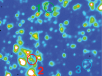 Photovoltaic cells - A new characterization method based on hyperspectral imaging recording spectrally resolved images allowing the cartography of electroluminescence and photoluminescence.
From the data acquired spatial variations of cell properties such as open circuit voltage and transport mechanisms were identified and characterized.
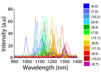 NIR Fluorescence of single nanotubes in live cells - IMA can be used for wide-field near-infrared hyperspectral imaging to spatially observe the fluorescence and spectral heterogeneity from single nanotube in living systems.
This approach resolved up to 17 distinct species (chiralities) with single nanotube spatial resolution in live mammalian cells, murine tissues ex vivo, and zebrafish endothelium in vivo.
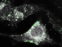 Nanoparticles in cancer cells - Photon etc.’s hyperspectral imager, IMA PL™, can be equipped with a highly efficient dark field condenser and generate high contrast images of biological samples like human breast cancer cells.
Contact Photon etc for more information on these applications
|
Technical Specifications
|
IMA PL VIS |
IMA PL SWIR |
| Spectral Range |
400 - 1000 nm |
900 - 1700 nm |
| Excitation Wavelength |
532 nm |
808 nm |
| Spectral Resolution |
< 2.5 nm |
< 4 nm |
| Spatial Resolution |
Sub-micron |
| Microscope |
Upright |
| Objectives |
20×, 50×, 100× |
| Maximum Sample Format |
4" x 4" (10 cm x 10 cm) |
| X, Y Travel Range |
76 mm x 52 mm |
| Z Stage Resolution |
1 µm |
| Maximum Scanning Speed |
150 ms |
| Wavelength Absolute Accuracy |
0.25 nm |
| Video Mode |
Megapixel camera for sample visualisation |
| Preprocessing |
Spatial filtering, statistical tools, spectrum extraction, data normalization, spectral calibration |
| Hyperspectral Data Format |
FITS, HDF5 |
| Single Image Data Format |
JPG, PNG, TIFF, CSV, PDF, SGV |
| Software |
Computer with PHySpec™ control and analysis software included |
| Dimensions |
˜ 40" x 30" x 30" (102 cm x 76 cm x 76 cm) |
| Weight |
˜ 80 Kg |
|
Upgrades
|
| Laser |
Additional excitation wavelengths available |
| Camera HI |
Back-illuminated camera |
N/A |
| EMCCD |
N/A |
| High Resolution Module |
N/A |
900-1700 nm, FWHM < 1 nm |
|
Photon etc's Global Imaging Technology