 Cryo-transmission electron microscopy captured numerous hairlike appendages radiating from the surface of this ultra-small bacteria cell. The scientists theorize the pili-like structures enable the cell to connect with other microbes and obtain life-giving resources. The scale bar is 100 nanometers. (Credit: Berkeley Lab)
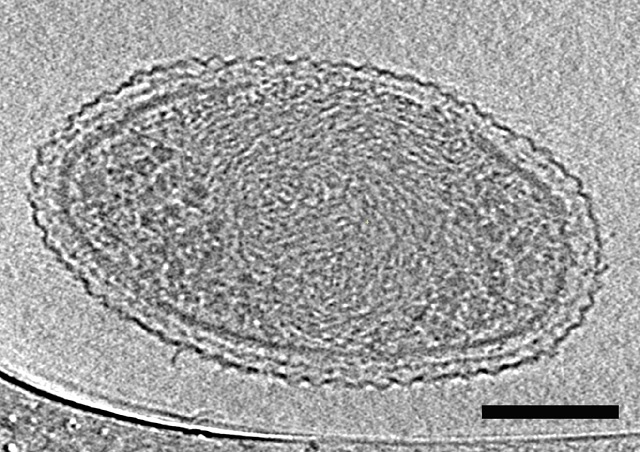
The existence of ultra-small bacteria has been debated for over two decades but until now there has not been a DNA-based description or comprehensive electron microscopy images of these microbes.
The average volume of the cells is just 0.009 cubic microns - around 150 of these bacteria would fit in an Escherichia coli cell and over 150,000 cells would fit on to the tip of a human hair.
This unique bacterium was discovered in ground water and it seems they are quite common. They are also strange, which is not surprising as the cells are very close together and in certain cases, smaller than a number of estimates for the lower size limit of life.
This is probably the smallest a cell can be and still have enough material to be a living cell. These bacterial cells contain few ribosomes, densely packed spirals which may be DNA, small hair-like structures and a stripped-down metabolism, which requires them to depend on other bacteria for the needs of life.
The bacteria belong to three least understood microbial phyla. The effect of microbes on the climate, water and food supply of our planet as well as other key mechanisms can be understood by studying about these bacteria from these phyla.
These newly described ultra-small bacteria are an example of a subset of the microbial life on earth that we know almost nothing about.
Jill Banfield, Senior Faculty Scientist, Berkeley Lab’s Earth Sciences Division
“They’re enigmatic. These bacteria are detected in many environments and they probably play important roles in microbial communities and ecosystems. But we don’t yet fully understand what these ultra-small bacteria do,” said Banfield.
Banfield is a co-author of the paper, which has been published in Nature Communications, along with Birgit Luef, a former postdoctoral researcher in Banfield’s group who is now at the Norwegian University of Science and Technology, Trondheim.
There isn’t a consensus over how small a free-living organism can be, and what the space optimization strategies may be for a cell at the lower size limit for life. Our research is a significant step in characterizing the size, shape, and internal structure of ultra-small cells.
Birgit Luef, Norwegian University of Science and Technology
The scientists began studying bacteria from phyla, which did not have cultivated representatives. Certain of these bacteria have very tiny genomes so the scientists concluded that the bacteria may also be very small.
Filtered groundwater collected at Rifle, Colorado, was subjected to several smaller filters, down to 0.2µm, in order to obtain a concentrated cell sample. 0.2µm is the size normally used for water sterilization.
The samples obtained were not sterile. They had very small microbes, which were subjected to flash freezing at -272˚C in a rare portable version of a device known as a cryo plunger. This made sure that the microbes were preserved even during transportation from the field to the lab.
The frozen samples were moved to Berkeley Lab, where Luef, along with Luis Comolli of Berkeley Lab’s Life Sciences Division, performed cell internal structure and size characterization with two-dimensional and three-dimensional cryogenic transmission electron microscopy. It was also observed that the cells were dividing, showing these were healthy bacteria and not in that ultra-size, because of starvation.
The sequencing of the genomes of the bacteria was done at the Joint Genome Institute, a DOE Office of Science User Facility located in Walnut Creek, California, under the guidance of Susannah Tringe. The length of the genomes was around one million base pairs. Furthermore, metagenomic and other DNA-based analyses of the samples were performed at UC Berkeley, which found several bacteria from WWE3, OP11, and OD1 phyla.
This integration of sophisticated microscopy, genomic study and excellent fieldwork helped obtain the most comprehensive description of ultra-small bacteria till date.
The researchers observed that certain bacteria had thread-like structures known as pili, which could act as life support connections to surrounding microbes. The genomic data showed that the bacteria lacked several basic functions as they depended on a microbe community for their survival.
The scientists also noted that there is a lot more to learn about ultra-small organisms.
We don’t know the function of half the genes we found in the organisms from these three phyla.
Jill Banfield, Senior Faculty Scientist, Berkeley Lab’s Earth Sciences Division
The Advanced Light Source, a DOE Office of Science User Facility located at Berkeley Lab, was also used for the study, where Hoi-Ying Holman of the Earth Sciences Division helped in determining that most of the cells in the samples were bacteria and not Archaea.
The study is a marked contribution to what is already known about ultra-small organisms. Scientists recently estimated that a marine bacterium’s cell volume is at 0.013 cubic microns;however, the method used did not determine the cell diameter directly.
There are also previous electron microscopy images of an Archaea lineage with cell volumes as tiny as 0.009 cubic microns, very much like these bacteria, which include results of work by certain of the same researchers.
Combined, the study findings show the existence of small cells with odd and considerably restricted metabolic capacities from two of the three key branches of the tree of life.
This research has been published in the journal Nature Communications.