Researchers at the University of Chicago are developing computer-aided diagnosis (CADx) and quantitative image analysis (QIA) methods for mammograms, ultrasounds and magnetic resonance images (MRIs) to identify specific tumor characteristics, including size, shape and sharpness, said lead researcher Maryellen Giger, A.N. Pritzker Professor of Radiology/Medical Physics and director of the Imaging Research Institute at the University of Chicago.
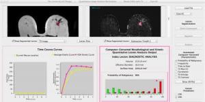 The quantitative image analysis workstation in the Giger laboratory for assessing breast lesions observed in MRIs, showing automated lesion segmentation, feature extraction (volumetrics, morphology, texture, kinetics), and estimation of the probability of malignancy. Credit: University of Chicago
Currently, computer-aided detection provides a "second opinion" to a radiologist in locating suspicious regions within mammograms. Next, radiologists will ultimately be able to use computer-extracted lesion characteristics when performing a diagnosis to assess whether the tumor is cancerous.
The role of quantitative image analysis is expanding beyond screening and toward application of risk assessment, diagnosis, prognosis, and response to therapy, and in using data to identify how tumor characteristics apply to disease states, Giger said.
This could lead to the comparison of a tumor's characteristics with thousands of similar cases, enabling the exploration of complex relationships among tumor characteristics across large populations, which may ultimately contribute to the design of patient-specific treatments. It could also be used to study the association between a tumor's observable characteristics and cell-level data for the emerging field of imaging and genomics, which aims to identify genes that influence the risk for disease.
While results are promising for digital mammograms, researchers are extending their analysis to breast ultrasounds and MRIs due to the need for clinical validation within a larger screening population.
Through studies between image-based characteristics and genomics, investigators will potentially be able to determine which tumor characteristics are related to and which complement genetic findings, with the ultimate goal of merging them to include both genetic and environmental contributions in clinical decisions. Researchers are now using data-mining methods to identify those potential relationships.
A paper titled "Quantitative breast image analysis for personalized medicine" describing the work by Giger was published 14 October in the SPIE Newsroom.
Source: http://spie.org/