A variety of advanced techniques have been developed to characterize the atherosclerotic plaque, including multidetector spiral computed tomography, magnetic resonance imaging, intravascular ultrasound, optical coherent tomography and intravascular near infrared spectroscopy.
Pu Wang and his colleagues from Purdue University, West Lafayette, and Indiana University School of Medicine (USA) now developed a promising new one: They employ an optical window between 1600 nm and 1850 nm for bond-selective deep tissue imaging. Label-free imaging of atherosclerotic plaques can be performed through optical excitation of first overtones of CH bonds and acoustic detection of the generated ultrasound waves in this previously underappreciated optical window.
Until now, the consensus was that the gold optical window lies between 650 and 1300 nm and that it stops at 1300 nm due to significant water absorption at longer wavelengths. The scientists noted that water absorption in the region beyond 1300 nm is modulated by the vibration transition of H2O: They observed a significant valley between 1600 and 1850 nm, where the absorption coefficient of pure water is at the same level as that of heme proteins in whole blood around 800 nm.
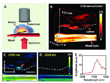
Considering the reduced scattering and diminished phototoxicity at longer wavelength excitation, the wavelength region from 1600 to 1850 nm is appealing as a new optical window for deep tissue imaging (light red shadow region). Importantly, the first overtone of CH vibration is located at the same window. Using this new window to carry out photoacoustic imaging, the scientists found a 5 times enhancement of photoacoustic signal by first overtone excitation of the methylene group CH2 at 1730 nm, compared to the second overtone excitation at 1210 nm.
This enhancement allowed 3D mapping of intramuscular fat with improved contrast and of lipid deposition inside an atherosclerotic artery wall in the presence of blood. Moreover, lipid and protein could be differentiated based on the first overtone absorption profiles of CH2 and methyl group CH2 in this window.
Selective vibrational photoacoustic microscopy imaging of collagen and lipids heralds the potential in diagnosis of vulnerable plaques through detection of the thickness of the collagen cap and the location of the lipid-laden plaque inside the arterial wall without molecular labeling that could alter tissue composition.
Source: Journal of Biophotonics