During and after operating procedures, surgeons use three-dimensional X-rays for performing checks. The three-dimensional images enable them to avoid the risk of complications, follow-up surgeries and also allow them to verify the position of fracture fragments and implants.
 The ORBIT 3D X-ray scanner makes it possible to capture X-ray images during an operation quickly and without time-consuming preparations. Courtesy: Fraunhofer Institute
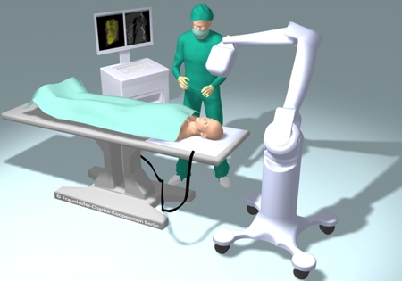
Existing C-arms and similar three-dimensional X-ray systems interrupt and interfere with the surgery. The detector as well as the X-ray source has to be moved in a circular manner around the patient. The equipment has to be moved to the operating table when required and then moved back. This would interrupt the surgery and also consume time. If the equipment was fixed on the operating table it would hinder the work of the surgeon.
Scientists at the Fraunhofer Institute for Production Systems and Design Technology IPK, Charité – Universitätsmedizin Berlin university hospital and Ziehm Imaging are jointly working on developing ORBIT, a three-dimensional X-ray scanner for capturing X-ray images during surgeries without any major interruptions. In the ORBIT system, the X-ray source moves in a circular path over the operating table. This avoids the time taken for preparations and it can also capture images quickly. The system does not have to surround the patient. The ease of use will enable surgeons to utilize the device regularly.
In C-arm scans, screws and implants may cause interference. But in the ORBIT system the X-ray detector and source do not move along the same plane, which leads to fewer artifacts in the images. The system consists of an X-ray source, a digital flat panel detector and a display monitor. The X-ray source is fit to an articulated bracket and can be maneuvered. The bracket can be mounted on a mobile stand or fixed to the ceiling. The detector is set in the operating table and the X-ray display monitor can be wall-mounted or kept mobile.
A prototype is under construction and testing is to commence in 2012. More information on ORBIT will be available at the Medica 2011 trade fair in Düsseldorf.