May 26 2017
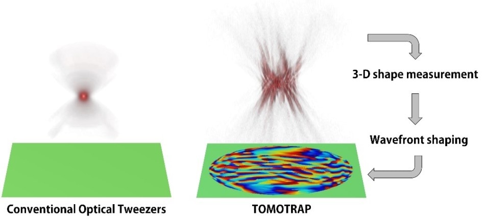 Concept of optical manipulation techniques. Credit: KAIST
An optical manipulation method with the ability to freely control the orientation, position, as well as shape of microscopic samples with complex shapes has been developed by Professor YongKeun Park and his team from the Department of Physics at KAIST. The research was published online on 22 May 2017 in the journal Nature Communications.
Traditional optical manipulation methods, also known as “optical tweezers,” have been applied as a crucial means for applying micro-scale force on microscopic particles to control the three-dimensional (3D) positions of the particles. Optical tweezers include a tightly-focused laser with a beam diameter less than 1 mm, or one-hundredth of thickness of the human hair, thus exerting attractive force on adjacent microscopic particles traveling toward the beam focus. By manipulating the positions of the beam focus, the research team was able to control the particles and move them freely to other positions, hence the name “optical tweezers.” Optical tweezers have been extensively applied in different areas of biological and physical research.
Until now, a majority of experiments carried out using optical tweezers have been performed for trapping spherical particles. This is because physical principles can estimate optical forces and the reciprocating motion of microspheres in a simple manner. However, in the case of trapping objects that have complicated shapes, traditional optical tweezers produce unstable motion of the particles, and controllable orientation of these objects is restricted, thereby hampering the regulation of 3D motion of microscopic objects that have complex shapes, for example, living cells.
The researchers created an innovative optical manipulation method with the ability to trap complex objects that have arbitrary shapes. This method involves primarily carrying out real-time measurement of 3D structures of an object by using a 3D holographic microscope. The physical principle of a 3D holographic microscope is same as that of X-Ray CT imaging. The researchers used the measured 3D shape of the object to accurately compute the shape of light that has the ability to steadily control the object. If the shape of the object and that of the light are similar, the object’s energy is reduced, thus enabling stable trapping of the object with the complex shape.
In addition, when the shape of light is controlled to have different orientations, positions, and shapes according to the objects, the 3D motion of the object can be freely controlled to produce an object with a desired shape. This procedure is similar to generating a mold to cast a statue with a desired shape, so the researchers termed this method “tomographic mold for optical trapping (TOMOTRAP).” The researchers were successful in stably trapping individual human red blood cells, rotating them to desired directions, folding them into L-shape, and arranging two red blood cells together to produce a new structure. Furthermore, they could also stably trap colon cancer cells that had a complex shape and rotate them to desired directions. These have been highly difficult to achieve by using the traditional optical methods.
Our technique has the advantage of controlling the 3-D motion of complex shaped objects without knowing prior information about their shape and optical characteristics, and can be applied in various fields including physics, optics, nanotechnology, and medical science.
Professor Yongkeun Park, Department of Physics, KAIST
The lead author of the paper, Dr Kyoohyun Kim, stated that the new method has the ability produce controlled deformation of biological cells that had desired shapes. “This approach can be also applied to real-time monitoring of surgical prognosis of cellular-level surgeries for capturing and deforming cells as well as subcellular organelles,” further stated Kim.
Controlling 3D Behavior of Biological Cells Using Laser Holographic Techniques